(Myths and Facts)
Article posted on the site November 11, 2013.
(Handbook for the Study toksoplazmid)
V.A.Bugaev, A.M.Bugaev
SUMMARY
Over many years of work on the book "Toxoplasmosis small pets - Myths and Facts." Authors - Bugaev Vladislav - President of the Association of Veterinarians of Kazakhstan and Bugaev Anatoly Makarovich - Director of the Center for Animal Health, Kiev.
The book summarizes the results of years of their own experimental research on the life cycle of Toxoplasma and their relationship with the host. Benefit illustrated many unique photos made by the authors. Photos demonstrate certain life cycle processes toksoplazmid, their morphology and parazitohozyaennye relationship. Based on the analysis of literature data and own research, the authors have tried to clarify some of the myths developed recently in the problem of toxoplasmosis. Paid great attention to the role of cats in the epizootiology and epidemiology of toxoplasmosis of humans and animals. Proofs are given the important role of Toxoplasma strains slabovirulentnyh impact on animals and especially cats during pregnancy. Discusses the position of Toxoplasma and other toksoplazmid the system simple. The importance of the book is devoted to the head of diagnosis and treatment of toxoplasmosis in small pets.
The book is intended primarily for medical and veterinary pathologists studying the life cycles of parasitic protozoa and the relationship with the host. The materials presented in the book are of some interest to biologists, parasitologists, university professors appropriate profile, as well as practicing veterinary and medical doctors.
Particular attention is paid to the terminology and toksoplazmidozah toxoplasmosis, as well as systematics toksoplazmid. So while negotiations are underway on the publication of this manual, we would like to discuss social networking, open press and on the forums, some of the chapters of this book - it questions relating to our proposed terminology, systematics toksoplazmid and answer the most important question - Toxoplasmosis in people - it Myth or Reality?
Our long experience shows that toxoplasmosis for animals - this is a real problem that must be studied and solved by improving methods of diagnosis and treatment of disease. Particularly toxoplasmosis, as the problem is important for animals that are kept in the home with them and have the closest contact. And the task of veterinarians, physicians discuss toxoplasmosis until a problem or not for people to minimize the contamination of people, keeping in mind that "Veterinary treats humanity." In addition, we must remember that animals - domestic and farm, found another protozoan disease caused by parasites, the life cycle and the impact on the animal organism which is similar to Toxoplasma - it NEOSPOROZ. Therefore, the solution of the problem of toxoplasmosis, will depend largely on the study and neosporoza.
Authors are grateful for the constructive criticism and will answer all your possible opponents and colleagues who really want to understand this overarching, daunting problem of human and veterinary medicine.
Sincerely V.A.Bugaev, and A.M.Bugaev
Tellefon Contact - (044) - 426 - 47 - 76 (78).
Email:
Этот e-mail адрес защищен от спам-ботов, для его просмотра у Вас должен быть включен Javascript
NOTE: All photos that do not give credit, owned AM Bugaev and V.A.Bugaevu and stored in our archive.
About C D E F G H A & E
CHAPTER ONE
PREDISLOVME
INTRODUCTION
TERMINOLOGY
CHAPTER TWO
GENERAL ISSUES IN THE NATURE OF CIRCULATION Toxoplasma - MORPHOLOGY, REPRODUCTION, SYSTEMATICS
1.TSIRKULYATSIYA Toxoplasma IN NATURE
2. MORPHOLOGY AND REPRODUCTION In toxoplasma
ORGANISM intermediate hosts
MYTH ONE. Toxoplasma reproduce only intracellularly.
MYTH TWO. In the body of the intermediate hosts of Toxoplasma reproduce only by endodiogenii and poliendogenii.
MYTH THREE. Pseudocysts of Toxoplasma tissue cysts turn into breeding.
3. EDUCATION CYSTS Toxoplasma gondii in the brain WHITE
MICE
4.OBRAZOVANIE CYSTS Toxoplasma gondii in skeletal
MUSCLES white mice
5.MORFOLOGIYA Toxoplasma gondii CATS IN THE BODY.
MYTH FOUR - when fed to cats raw meat infested Toxoplasma in their gut parasites necessarily pass through the phase of sexual development and reproduction.
6. Toxoplasma MORPHOLOGY IN EPITHELIAL
Intestinal cells CATS
7. MORPHOLOGY VNEEPITELIALNІH toxoplasma In
GUT CATS
8. SISTEMATIKA toxoplasma
The fifth myth. Toxoplasma gondii - is coccidia.
CHAPTER THREE
PARAZITOHOZYAINNYE RELATIONSHIP WITH
EXPERIMENTAL ANIMALS toxoplasmosis
MORPHOLOGICAL CHARACTERISTICS OF PRIMARY FOCUS IN
Toxoplasmosis
1. Morphological characteristics of the primary lesion
serous membranes in albino mice at vnutribryu-
tire infection with a virulent strain CDN
2. Morphological characteristics of the primary lesion at the site of intramuscular injection of a virulent strain of Toxoplasma RH
3. Morphological characteristics of the primary lesion at the site of intramuscular injection of Toxoplasma strains slabovirulentnogo LEI.
4. Morphological characteristics of the primary lesion at the site of intramuscular injection izospor slabovirulentogo strain of Toxoplasma LEI.
5. Morphological characteristics of the primary lesion
on the site of intracerebral administration to mice izospor
Toxoplasma strains slabovirulentnogo LEI
6. Morphological characteristics of the primary lesion in the intestine at the site of introduction of the virulent Toxoplasma
strain RH
7. Morphological characteristics of the primary lesion after intraperitoneal infection of Toxoplasma puppies
virulent CDN
General conclusion on pathomorphological characteristics of primary toxoplasmosis FOCUS IN DIFFERENT WAYS OF ADMINISTRATION toxoplasma
CHAPTER FOUR
MORPHOLOGICAL CHANGES IN ORGANS OF ANIMALS
IN EXPERIMENTAL toxoplasmosis
1. WHEN acute toxoplasmosis
2. Subacute toxoplasmosis
3. WHEN chronic toxoplasmosis
4. In latent toxoplasmosis
MORPHOLOGICAL CHANGES IN THE BRAIN
PILOT toxoplasmosis
The fifth myth. Slabovirulentnye strains of Toxoplasma are not dangerous to humans and animals.
CHAPTER FIVE
Toxoplasmosis in DIAGNOSTICS CATS AND DOGS
CHAPTER SIX
Treatment of toxoplasmosis in cats and dogs
1. CHRONIC toxoplasmosis
2. TOXOPLASMOSIS In latent
INTRODUCTION
Following the publication of the article "Toxoplasmosis. A new look at an old problem "in the journal New medical technologies number 4 and 5, 2005. Eight years have passed. Over the years the material in this article were perekopirovat on different sites and, unfortunately, with large discrepancies. This is especially true numbering photos and captions. Although these errors are made through no fault of the author and editors of the journal, we apologize to readers for certainly interesting from a scientific point of view the material, but in such an ugly form submitted to sites on the Internet.
Glory to God! In recent years, the problem of toxoplasmosis comes from "oblivion." Close it and failed, despite the efforts of AY Lysenko and his disciples. Finally life has put everything in its place. Not only supporters but also the opponents of toxoplasmosis problems appeared vital need for a practical use of previously accumulated knowledge, both in diagnosis and treatment of toxoplasmosis in humans and animals. In the years to discredit the researchers and the total closure of laboratories have studied the problem of toxoplasmosis in the USSR, the first researcher and organizer of the study of toxoplasmosis in the Soviet Union, Professor Dmitri Nikolaevich Zasukhin to support their students and colleagues said: "Close the problem of toxoplasmosis is impossible, as it is impossible to close an open America "- quoted by EA Shevkunova (2006).
Confirmation of these prophetic words of the scientist is the emergence in recent years in Europe, America and the CIS, many capital works setting forth the unique factual material as to the clinic, diagnosis and treatment of toxoplasmosis and toxoplasmosis parazitohozyaennym relationship. And although some of the facts today sometimes still difficult to explain, but they could no longer ignore, much less ignored. We stand on the threshold of new hypotheses and interpreting the resulting new factual material as the problem of toxoplasmosis and neosporoza. This is particularly true of so-called latent or "bessimtomnogo" of the disease, which some doctors take as innocuous form flow toxoplasmosis. Academician O. Yiorovets from Czechoslovakia (1962), Professor LK Korovitskiy (1966) and Professor A.P Kazantsev (1985) from the Soviet Union, the first to draw attention to this phase of toxoplasmosis flow and gave a precise definition of this stage of the disease - "PSEVDOLATENTNAYA."
I would like also to draw the readers' attention on these issues in publications that come out of the leading centers and individual researchers to study the problem of toxoplasmosis - Works of the Russian Navy - Medical Academy, St. Petersburg; Toxoplasmic Odessa regional center, as well as work on OI Eleseevoy, especially its article - "Toxoplasmosis, a technique refined diagnosis in vegetative resonance test"; publication and presentation Ukrainian doctors - Dr. Krivonosa Vladimir Kutuzov and Dr. EO Kamarouski. On the Interpretation of some of the issues Dr. E.O.Komarovskim toxoplasmosis problem is debatable. However, one must give him credit for that by using the latest technical possibilities televidniya, Internet and Yutuba he did more than any researcher, in terms of understanding the problem and prevent the disease toxoplasmosis.
If the CIS scientists, medical and veterinary doctors "restore" the problem of toxoplasmosis from the ruins, in the west of the country study of this problem has never ceased. At the time when the Soviet Union in the late 20th century closed laboratory for the study of toxoplasmosis and we were drawn into an unnecessary debate - "Is it important or not important problem for toxoplasmosis nashogo public", "Toxoplasma Coccidia Coccidia or not", "Who is the leader in this problem, "Western scholars have set ourselves the task, as better and without complications to treat people and animals sick with toxoplasmosis. And in this respect, in our opinion, have achieved great success employees Berlin Robert Koch Institute, under the leadership of professor Frank Sieber (Frank Seeber) conducted a unique work to find new chemical medicines to treat patients with toxoplasmosis. It is very important that they pay attention to the common morphological structures of Toxoplasma and malaria parasites (united us in one subclass Haemosporina), acting on which researchers hope lichit not only toxoplasmosis, but also malaria.
The basis of this book, the authors have put research Veterinary Clinic Center for Animal Health Dr. AM Bugaev Kiev and the Veterinary Diagnostic Center Vladislav Bugaev Almaty. The paper also benefited professor V.F.Novinskoy and SNA UD WUSTIN - toxoplasmosis laboratory researchers of the Institute of Zoology. Kaz. SSR. which in combination with AM Bugaev worked from 1964 to 1977 under the direct supervision of academician Hilarion G. Galouzeau and professor, department chairman patanatomii farm animals Almaty Red Banner of Labor and Veterinary Institute Konstantinov Pavlov Vsevolodova.
Much of the material included in the book has already been published in various publications of the last century and in the book "The life cycle of Toxoplasma" which was published in 1974 under the editorship of I.G.Galuzo, as well as presented at the Third International Congress Protozoologists in 1969. Some of the material is taken from the doctoral dissertation AM Bugaev and published. Given that in the book "The life cycle of Toxoplasma" of - for the technical possibilities of the time, the illustrations for the works were of low quality, we have tried to correct technical errors that time and present illustrative material in a form in which it is stored in our archives. We hope that the present paper will allow doctors to take a new look not only at the illustrative material, but also on the interpretation of it, and thereby help to rethink some issues problems toxoplasmosis. Since the problem of toxoplasmosis more and more reminds himself not only in terms of the reproduction of a healthy population of the planet, but also through blood transfusions, organ transplants, surrogate motherhood, HIV - infected people and in general for any immunosuppression. More and more publications appears on the relationship of schizophrenia with Toxoplasma. Here's what we found about this in the materials - Wikipedia, the free encyclopedia - "Based on the latest scientific research it is believed that Toxoplasma gondii can provoke the development of schizophrenia [2] [3] [4] In the period since 1953 was undertaken by 19 research detection of antibodies to T. gondii in patients with schizophrenia, 18 of which had shown a higher incidence of HIV seropositive individuals with mental disorders, and in 11 studies the results were statistically significant (p <0.05). Thus, in one study, antibodies were detected in 495 (52%) of 961 patients with schizophrenia compared with 170 (25%) of 681 in the control group; else - at 836 (86%) of 973 patients with schizophrenia in comparison with 30% in the general population. It has also been shown to reduce the level of antibodies in patients treated for schizophrenia, compared with nepoluchaetsja, which is consistent with the data on the activity of certain antitoksoplazmoznoy antipsychotics. "
Remembering the work in the laboratory of the Institute of Zoology of toxoplasmosis. Kaz. SSR. performed under the leadership of I.G.Galuzo, you know how important and unique large-scale studies conducted in this laboratory. At the moment, it is difficult to imagine that what - either laboratory or institution in the CIS will conduct similar research. No exaggeration to say that such studies providence could only afford Great Country - Country that when - then called the Soviet Union, with its scientific and material potential. Therefore, published in the archival material in which - it least, are unique in that they repeat them in the near future, practically, it will be impossible or financially or technically.
The authors believe that the work will also help veterinary practitioners better understand the problem of toxoplasmosis and will improve as the diagnosis and treatment of toxoplasmosis in small pets. Since all animals infected with Toxoplasma, in one way or another, are dangerous to humans.
In toksoplazmid problem and in particular toxoplasmosis human and animal remains much more unexplained mysteries. Therefore, the authors hope that thoughtful medical doctors, as well convinced of the need for joint efforts of veterinary and medical professionals, the new rethink the entire accumulated in recent years, data on human and animal toxoplasmosis. And the sooner it happens the more successful will be solved the problem of toxoplasmosis, both in medicine and veterinary medicine. A writing and acting on television and on YouTube physicians, we advise first read and become familiar with the book of professor Ellen Anatolevny Shevkunova - "History of the study of toxoplasmosis with the Soviet Union - Chronicle research, memoirs, letters." E.A.Shevkunova is one of the first of many students prfessora Dmitry Nikolayevich Zasukhina - Organizers of the first with the USSR laboratory toxoplasmosis. Many of the provisions on the issue tosoplazmoza outlines the work is not lost relevance in the present time.
INTRODUCTION
TOXOPLASMOSIS - the most common natural - patchy, a parasitic disease of humans and animals (including poultry and wild birds), to conquer all the continents of the world. Infected people in different countries ranges from 10 -15 to 80% of animals from 5 to 6 to 60%. Almost every third person on the planet is infected with Toxoplasma, which has led some doctors believe that Toxoplasma safe for humans and animals.
Toxoplasmosis is caused by a single-celled parasitic protozoan Toxoplasma gondii. Morphology and endogenous Toxoplasma their breeding cycle is very similar to the malaria parasite, Leishmania, undeniable, Thaler and other blood parasites. Although Toxoplasma is simply called, but in its organization and life cycle they are so complex that over the years caused a lively discussion. And on the effects on humans and animals, Toxoplasma among the most insidious pathogens that are known to date medical and veterinary doctors as strike in the first place, the brain of humans and animals, causing significant economic losses to livestock, as well as affect fertility healthy population.
Despite the fact that in the last century shestidisyatyh opened sexual phase of reproduction in Toxoplasma, which are parasites in the intestines of cats (Hutchison, Work, 1969; Work, Hutchison, Siim, 1969 Galouzeau, 1970), toxoplasmosis and to date remains one of the most mysterious human and animal diseases around which flare up from time to time difficult debate. One such debate arose in the late twentieth century, the consequences of which have postponed mark on the consciousness of an entire generation of medical and veterinary doctors. Had already progressed to the point that part of medical and veterinary doctors have begun to argue that toxoplasmosis is not much - any significant problem for human and veterinary medicine.
Although reliable data of those who for many years worked on the problem of toxoplasmosis and every day meets people infected with Toxoplasma show otherwise. Thus, VV Vasiliev Vasiliev IS, 2009, from St. Petersburg in the article "Toxoplasmosis: modern scientific and practical approaches
Part X. Toxoplasmosis in beremennyh1 and congenital toxoplasmosis "analyzing the data reported in world literature," that the frequency of infection in pregnant women in different regions of the world, depending on the applied methods and test systems ranges from 22% (Israel) to 83.5% (Madagascar) averaging about 40% in average 1% of women are infected for the first time during pregnancy. "
A I.V.Lipkovskaya article "Clinical and pathogenetic aspects of toxoplasmosis," published in the early twenty-first century (2000 the) analyzing the data obtained in the center of the Odessa Regional toxoplasmosis writes: - "draw attention to themselves, which in recent years has increased the number of infected infants, especially in unscreened and untreated mothers before conception. From applying to the 1999 - 2000 in the center of Odessa toxoplasmosis, chronic toxoplasmosis confirmed by clinical observations and laboratory tests in 240 women of childbearing age. " A long time ago, is scientifically proven that a great threat especially toxoplasmosis is specifically for pregnant women and children. Professor AP Kazantsev (1985) believes that transplacental transmission of Toxoplasma to the fetus can lead to premature birth, fetal death, the development of his deafness, blindness, the backlog in the psychophysical development, cerebral palsy, hydrocephalus, etc. According to him infection of the fetus can occur in 60% cases, if the mother did not receive appropriate treatment in a timely manner. And most importantly AP Kazantsev (1985) drew particular attention to the fact that the initial development to recognize symptoms of the disease, as well as to predict the outcome of labor and further development of the unborn child, is almost impossible. A well-known American scholar Jack Remington (GSRemington, 1990.) For its research, practice, confirms the findings of Professor AP Kazantsev. He is long, for over 9 years, watching a group of children with primary congenital latent invasion, concluded that 85% of these children developed late manifestations of congenital toxoplasmosis - encephalitis, convulsive disorder, mental retardation and eye damage. Based on my personal observation, Jack Remington believes that among children born to mothers who infested slabovirulentnymi Toxoplasma strains may be children, both healthy and sick. And clinically manifest, the disease can only later during the first 8-9 years of life.
In a review article I.V.Lipkovskoy (2000) also states that sometimes signs of the fetus may be so mild that at birth the child seems to be quite healthy. Intrauterine lesion manifests itself later in the form of chorioretinitis, mental retardation, hearing loss or other defects in the nervous system.
According to the observations is explicitly I.V.Lipkovskoy rejuvenation disease. The largest number of people seeking help in the Odessa Regional Center of toxoplasmosis, were persons older school-aged and younger - 12 to 44 years - 85%. The author also notes the increase in the number of exacerbations and relapses of toxoplasmosis in previously untreated individuals with relatively good general condition. Therefore, as the facts show, the absence of overt clinical signs at toksoplazmonositelstve - seropositive or having only immunoglobulin G, is not yet a sign of health and especially safety.
After the opening stage of sexual reproduction in an organism Toxoplasma felines, all the troubles with Toxoplasma infection of people practically shifted to domestic cats. And many doctors advise women before planning pregnancy and during pregnancy to get rid of the cats. And as shown by our experience and practical data of numerous studies in many countries, domestic cat that does not get raw meat and is not walks by itself is not a source of human infection with toxoplasmosis. It is only necessary to organize the mode of keeping and feeding the cats, as well as check on the cat and the cat toksoplazmonositelstvo. No less dangerous in terms of human infection with toxoplasmosis are other pets, especially canines, which are fed raw meat.
More significantly in the last century it has been proven that the use of raw, poorly thermally processed meat is the main route of human infection. Despite this, even in the most civilized countries, you can apply in the restaurant's signature dish - steak with blood and other meat dishes with meat neprozharennoe. Many forget that mice and rats are no less dangerous for the spread of Toxoplasma in nature than the cat being in grocery stores and warehouses, as well as in closed reservoirs inhabited by rodents and amphibians, and reptiles, are ill with toxoplasmosis . The latter can also be hosts for Toxoplasma transit (A.V.Levit, 1974). Therefore, the problem of toxoplasmosis for veterinarians is no less important than for physicians, both in terms of maintaining the health of small domestic animals, and in terms of prevention of human disease. In addressing the problem of toxoplasmosis, veterinarians must come not only from the perspective of the treatment of domestic animals, but also to avoid contamination of people as the main source of human infection are livestock and pets. In this regard, practicing veterinarians, at the present stage of our knowledge of the morphology and biology of Toxoplasma, their influence on the animal organism, it is necessary to have a scientific basis:
1. Correctly interpreting the readings of diagnostic tests, and first of all serological tests, ELISA and PCR;
2. For an informed decision whether or not to treat animals when they detected antibody class G;
3. Effective and gentle treatment of toxoplasmosis in animals;
4. Control, the effectiveness of the treatment of animals.
On these issues, the community of veterinary practitioners, and medical doctors in the CIS countries, divided into two camps. Some believe that the problems do not exist toxoplasmosis. It heirs and successors "ideas" Andrei Lysenko Jakovljevic, braked a case study of toxoplasmosis in the USSR for 15 years - 20. Second - doctors, occurring every day with this disease, wondered why instead of a deep study of the problem, with the use of modern methods of investigation, we are within more than 20 years, all discussing about the importance of toxoplasmosis for human and veterinary medicine. Many medical and veterinary doctors because of misinformation has even believe that the problem of toxoplasmosis and contrived this disease do not need too much to ignore.
Already comes to the point of absurdity, when doctors do not vidivshie never set eyes or Toxoplasma or toxoplasmosis sick people and animals, assigns the right to assert that toxoplasmosis is not a dangerous disease, like as ARI and that human and animal organism itself quite easily handles with Toxoplasma. A single most "advanced experts" even believe in happiness when people Toxoplasma infection occurs in early childhood. And the girls' parents! Advise quickly bring a cat into the house, so that she, as soon as possible Toxoplasma infected child, then they say, she (the girl) will eventually become a full-fledged mother. Just think about these tips!
To better understand the origins of this thinking, it is necessary to go back to the end of 70 and beginning of 80 years of the last century, passed away when prominent scholars of the late twentieth century - protozoology Professor Dmitri Zasukhin, founder of the first Soviet toxoplasmosis laboratory at the Institute of Epidemiology and Microbiology. Gamal AMS USSR and academician Hilarion G. Galouzeau, founder of the first laboratory of toxoplasmosis in Kazakhstan at the Institute of Zoology AN.Kaz, SSR., And the head of the problem of toxoplasmosis in the USSR became a professor, head of the USSR Ministry parasitologist Andrey Yakovlevich Lysenko. This is despite its enormous "organizing" the possibilities and abilities, in the period from 1977 to 1978 was closed in Moscow is one of the leading laboratories in the USSR - toxoplasmosis laboratory in the Institute. Gamal. And then, in practice, have been discontinued studies on toxoplasmosis and other cities of the USSR, where there were centers for the study of toxoplasmosis. This is his "light" hand print several "capital" works on toxoplasmosis stated that "toxoplasmosis - an extremely rare disease, almost always asymptomatic carriage, so toxoplasmosis should be treated as a healthy carriage ..." - quoted by E.A.Shevkunovoy ( 2006), History of the study of toxoplasmosis in the Soviet Union.
Thanks to the book Professor E.A.Shevkunovoy, we now know for where "grow legs" Discussion "asymptomatic" carriers of toxoplasmosis; of rarely expressed clinically acquired toxoplasmosis; and that treatment be only women infected during pregnancy and so on. " The only pity is that this protracted absolutely unnecessary discussion, CIS has grown for a new generation of medical and veterinary doctors finally entangled in this really very complex problem.
It is a pity that the "easy" hands AY Lysenko was closed the first Soviet laboratory toxoplasmosis, which was the National Center for the Study of toxoplasmosis in the USSR, that it "easy" hands, professor, devoted his life to studying the problem of toxoplasmosis, a renowned scholar, Dmitry Nikolayevich Zasukhin failed for "competition" and was dismissed from the laboratory, which was created. Contemporaries, who knew Dmitry Nikolayevich Zasukhina and Hilarion G. Galouzeau, and indeed to any sensible person, very difficult to understand how, for example, managed to AJ Lysenko, the third All-Union Symposium on toxoplasmosis, held in 1988 in Novosibirsk, even to reject the proposal of the Symposium organizers commemorate deceased eminent scientists of the twentieth century, Professor Dmitry Nikolayevich Zasukhina and academician G. Galouzeau Hilarion, who made a great contribution to the study of toxoplasmosis.
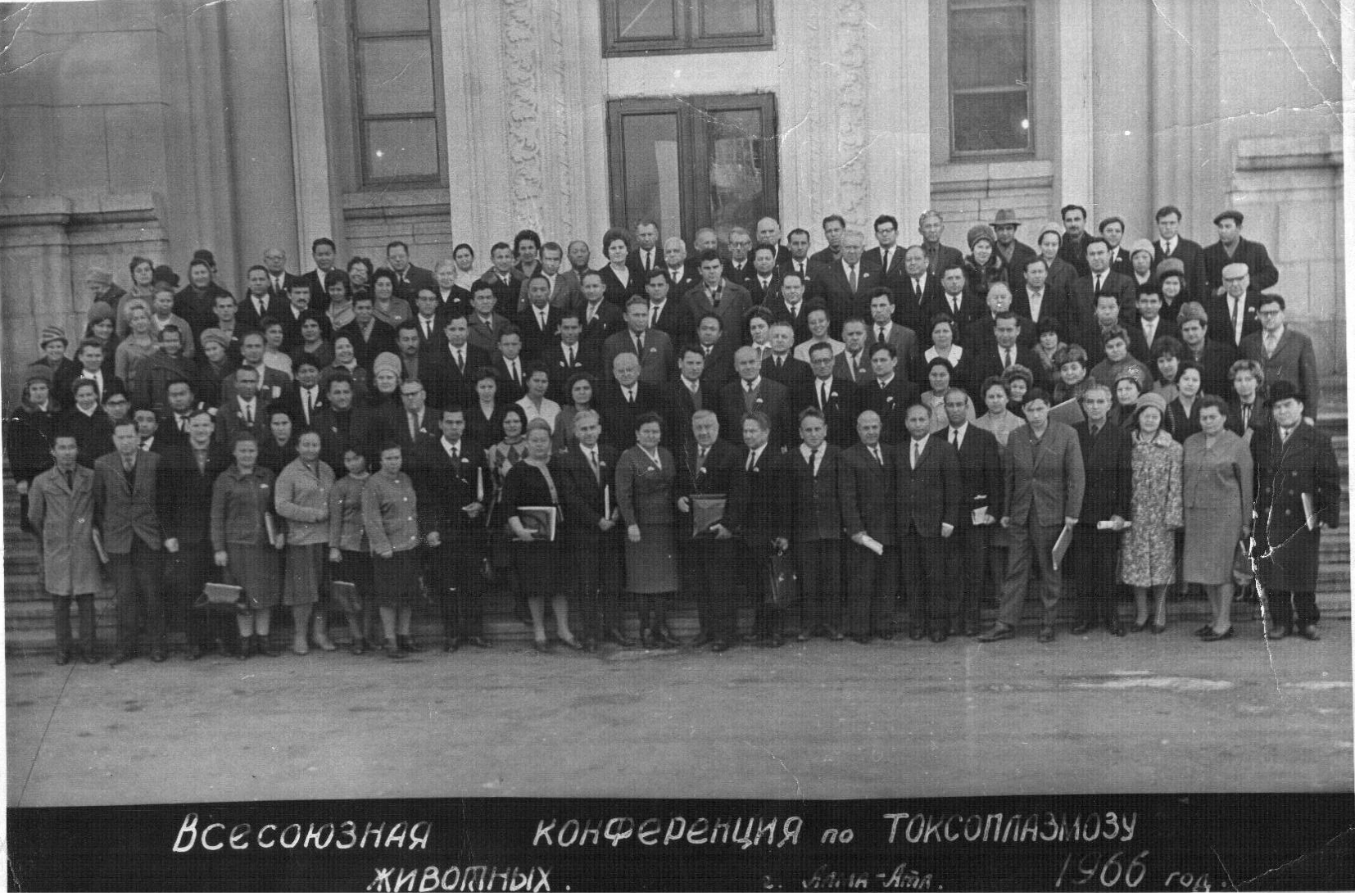
On the above photograph below shows the medical and veterinary doctors, as well as researchers, members of All-Union Scientific Conference on toxoplasmosis, held under the leadership of Hilarion G. Galouzeau and Professor Dmitry Nikolayevich Zasukhina in Alma-Ata in 1966. Many of the participants in the following years made a great contribution to the study of toxoplasmosis, became candidates, doctors and professors. Looking at the picture, one might wonder how one man managed to the end of the twentieth century, to close one of the leading laboratories for the study of toxoplasmosis in the USSR collapse and widespread conduct of scientific and practical work throughout the USSR?
NOTE. Not to be confused Academician Trofim Denisovich Lysenko, delay the development of genetics in the Soviet Union for several decades, Professor, Deputy Minister of Health (the late twentieth century) Andrei Yakovlevich Lysenko, braked a case study of toxoplasmosis in the USSR and CIS countries for two decades and has made a confusion in the heads of young medical and veterinary doctors early twenty-first century.
However, despite all the trouble that befell the Soviet scientists who have studied the problem of toxoplasmosis in 70 - 80 years, life has shown that the problem of toxoplasmosis in veterinary and human medicine there is, it is relevant and waits for its decision final. And it is very encouraging that in the last decade, the problem of toxoplasmosis visible shift from purely morphological study - biological issues to solve practical problems and veterinary medicine. Of the latter, capital works relating to the application of particular note veterinary doctorate Koroleva Svetlana Nikovaevna performed in 2003. In his literary analysis, it has once again reminded and confirmed how urgent is the problem of toxoplasmosis for veterinarians and physicians. Just think about the numbers! In the different countries of the CIS from 15 to 35% of cattle, sheep and pigs infected with Toxoplasma. And in foreign countries, according to her information, the percentage of animals seropositive for toxoplasmosis varies: in Italy antibodies to Toxoplasma gondii were found in 92% of cattle (F. Avezza et al., 1993), in France - 69% (A. Ca -bannes et al., 1997), in Greece, Spain - 40% (T. Moreno et al., 1991; M. Kritsepi-Konstantinou, 1992), in Brazil - 26% (JL Garcia et al., 1999) India, Turkey - 9% (M. Hafeez et al., 1992; N. Kucukerden et al., 1994), Norway - 5% (E. Skjerve et al., 1996). Toxoplasmosis among pigs registered in different countries: Italy - 64% (G. Genchi et al., 1991), Poland - 36% (M. Bartoszcze et al., 1991), Mexico, Canada - 9% (Z. Garcia-Vazquez et al., 1993; AA Gajadhar, 1998), Norway - 3% (V. Hirvela-Koski, 1992; E. Skjerve et al., 1996). In sheep antibodies to Toxoplasma identified France - 92% (A. Cabannes et al., 1997), Canada - 58% (D. Waltner-Toews et al., 1991), Brazil - 52% (JL Garcia et al., 1999), Mexico - 30% (Z. Garcia-Vazquez et al., 1990), Germany -21% (T. Gorman et al., 1999), China - 7% (DS Huang, 1991). Such a state of livestock, in epidemiological terms, ten times more dangerous than domestic cats, whose infection is only 3 - 5 percent. On the basis of their own data and analysis of the literature S.N.Koroleva once again showed that high rates of infection Toxoplasma animals and humans, primarily associated with a variety of ways of transmission. As well as climatic and sanitary conditions, especially ethnic cuisine, and the overall culture of the people.
The relevance of the problem of toxoplasmosis show the same author cited literature data on Toxoplasma infection of humans. In Moscow, Orel, and Omsk regions, identified respectively 25, 32 and 14% seropositive for toxoplasmosis people (LI Grachev, D. Goncharov, 1996). In Western Europe and North America are positive for toxoplasmosis, on average 25-50% to population, Africa, Central and South America - up to 90%. And the most dangerous thing that a high percentage toksoplazmonositelstva observed in women of childbearing age: Brazil - 72% (LC Rey et al., 1999), France - 54% (T. Ancelle et al., 1996), Germany - 39% (T. Beringer, 1992), Italy - 23% (A. Russo et al., 1999) Pakistan - 11% (MUAhmed et al., 1997), China - 4% (JH Hou et al., 1997). At the same time, medicine remains an open question to be treated or not women when it detects them high antibody class G.
Our many years of research and practical experience with small pets shows that unpredictable disease toxoplasmosis. And most importantly, all animals infected with Toxoplasma, in greater or lesser extent, dangerous to others, including humans. Therefore, we believe that all animals react positively to toxoplasmosis, regardless of whether they have identified IgM or IgG subject to compulsory treatment. Learn why we propose mandatory treatment of infected animals, you can read our book.
TERMINOLOGY for discussion
Reading and analyzing publications veterinary sites and forums, and not only veterinary unwittingly come to the conclusion that many writers on toxoplasmosis, clearly do not represent the whole problem. So often confused about the terminology stages of development and reproduction of Toxoplasma. This leads to the fact that it is sometimes difficult even for an expert to understand what the author wishes to express the idea in relation to pathologic process, or what stage of development and reproduction of the parasite in question. As an example confirming the above, an excerpt from a pretty serious work Valery Vasilyev (Department of Infectious Diseases of the Russian Navy - Medical Academy), who in his work Toxoplasmosis: modern scientific and practical approaches, Part Two. The unique properties of Toxoplasma. Life cycle and its clinical significance, "writes:" ... As mentioned in the first publication, Toxoplasma exist in three main forms: oocysts, tachyzoites and cysts. Subsequent form (cysts) itself is not a form of Toxoplasma, and is the formation of the walls composed of cells of the host organism, within which is a significant number of so-called bradyzoites. Apparently, readers are already completely confused (he writes) that is the same cysts and pseudocysts and how they differ from each other. I'll try to bring some clarity share. In modern foreign literature parasitological no term "pseudocysts" and there are oocysts, tachyzoites and bradyzoites (last - they - cysts). Differentiation of cysts and pseudocysts was introduced in the 40 - 50 years of the twentieth century pathologists that the term "cyst" understood necrosis (microabscesses) formed in the tissues as a result of multiplication of Toxoplasma, followed by the formation of dense granulation shaft consisting of immunocompetent cells. Inside the hearth no host cells, and contain multiple (5 - 10 thousand and more) bradyzoites. From the perspective of developing the disease cysts (those mikroabtssessy) - the final stage of development of relations with host cells Toxoplasma, which is realized under the scheme "tachyzoite" - "bradizoit" - "pseudocysts" (cyst) - "cyst" (mikkroabstsess). "
And now we ask (Authors), you dear readers understand after such authoritative clarification that such cysts or pseudocysts? We frankly can say that do not understand what the author wanted to express here and what does necrosis, microabscesses and granulating shaft of immunocompetent cells to have cysts or pseudocysts? Without entering into the debate here (it will be later) I would like to remind the author that even a novice pathologist knows what death is and what micro-abscesses and granulation tissue. And we believe that self-respecting pathologist, never take for Toxoplasma cyst necrosis or microabscesses. Therefore, if there are such grief - publications, they are not written clearly pathologists and they should be treated critically, especially in iternete published.
However we would not pay attention to the above rendition by cysts and pseudocysts, if the work Vladimir Vasiliev would not have been a really interesting and important in terms of practical solutions to issues of diagnosis and treatment of toxoplasmosis. And most importantly, if she did not come from the world-famous department, practically, one of the research centers not only the problems of toxoplasmosis, but also other natural - origin of disease, when - then headed by world-renowned scientist, Academician Yevgeny Nikanorovich Pavlovsky.
Yes, the problem toksoplazmid particularly toxoplasmosis, very complex, both in terms of pathogenesis and treatment, and in terms of endogenous and exogenous reproductive parasites. So that there is no verbiage in describing life cycles toksoplazmid and their relationship with the host, requires clear and precise formulation described stages of the parasite and resulting morphological changes.
In the study of any disease, after describing his pathogen development cycle, the relationship with the host and pathogenesis, there is its own specific terminology and concepts. And this is natural. This became particularly exemplified by studying the problem of toxoplasmosis and other toksoplazmid. For example, the concepts and definitions that are recognized by other parasitic protozoa, often do not reflect the fact toksoplazmidozah process or stages of the parasite, and practically not acceptable for toksoplazmid. And the use of toxoplasmosis such terms as "cyst coccidia", "classical and nonclassical coccidia", "typical and atypical coccidia", "tissue coccidia", "obligate - geteroksennye cyst coccidia", "tissue cyst eymereidnye coccidia" only make confusion in the description Toxoplasma and presentation of various aspects of the problem of toxoplasmosis. Therefore, the sooner such terms and definitions will disappear from our practice, the more successful it will address issues like toxoplasmosis and drugigih toksoplazmid.
In our special discussion vzlyad also requires writing in dogs and cats writing diagnosis - TSISTOIZOSPOROZ. Internet is full of such diagnoses. As evidenced this diagnosis? In - First, if in these cases we are talking about the diseases caused by Isospora felis and Isospora rivolta, or other izospor, the international call nosological terminology prescribes disease on the basis of the generic name of agent or historical names of the disease. If we adhere to the international terminology, the disease caused by I. felis and I. rivolta should be named and isosporiasis nikakakogo "TSISTOIZOSPOROZA" because no one offered to date classification serious parasitic protozoa of the genus CISTOISOSPORA not exist. At least, this is a generic name not universally recognized as such in the systematics and toxoplasmosis should be called neosporoz tsistoizosporozami. That in itself is absurd.
In - the second, all the studied species izospor significantly, in the body of intermediate hosts do not reproduce and do not form tissue cysts reproduction similar to those of toksoplazmid (Toxoplasma, Besnoitia, Sarcocystis, Neospora, Frenkelia, Hammondia). Rodents, for example, I. felis or I. rivolta are optional hosts to which they are phylogenetically not adapted. These parasites in the body intermediate hosts form resembling education disputes, and not reproduction cysts. In its time H. Mehlhorn and M. Markus (1976) called dormant stages or steps izospor expectations or dormozoitami but not cysts. Some researchers call them monozoitnymi cysts or hypnozoites. On the structure and biological function they correspond to steps sporozoites simple and not bradyzoites or trophozoites toksoplazmid. So call isosporiasis tsistoizosporozom makes no sense, because the prefix "cystitis" reflects nothing and generally meaningless.
If we are talking about, or rather on sarcosporidiosis sarkotsistoze, neither tsistoizospor nor izospor in the feces of a dog or cat, you often do not find, as in the external environment with the feces are not at izospory and sporocysts, so the prefix "cystitis" and "Arts "here is just not appropriate. And if we are talking about toxoplasmosis "tsistoizosporah" or undeniable, for them there are other nozoologicheskaya, all accepted scientific terminology is based on the generic name of parasites. It would be foolish, for example, will look like a doctor who obnatuzhiv in cat feces or zigotsisty izospory Toxoplasma, in the diagnosis of toxoplasmosis tsistoizosporoz write. We believe that our current level of knowledge about toksoplazmidah and in particular toxoplasma and toxoplasmosis, it is time to clarify the terminology and use terms that reflect the stage of development of parasites as well as their relationship with the host. We propose to discuss social networking, forums and in the press for toksoplazmid terminology listed below.
1. Toksoplazmidy - is strictly limited group toksoplazmopodobnyh parasitic protozoa , (Toxoplasma, Besnoitia, Sarcocystis, Neospora, Frenkelia, Hammondia),which are primarily parasites of leukocytes, and then RES cells and other tissues. In generalization phase and the chronic course of the disease, as toksoplazmidy able to multiply intracellularly and extracellularly. Some toksoplazmidy like sarkosporidii and Toxoplasma, and can invade erythrocytes (Photo 1 -8).
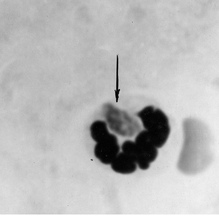
Picture 1. Plate preparation mesentery white mouse 2 hours after intraperitoneal inoculation of virulent strain of Toxoplasma CDN. Shows a well-formed trophozoites
T. gondii in the cytoplasm of neutrophils. Colouring Romanovsky - Giemsa.
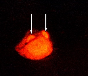
Photo 2. Smear prepared from abdominal exudate white mice experimentally infected with a virulent strain of Toxoplasma trophozoites CDN. Fluorescence microscopy. Drug treated akredinovym orange. Shows two trophozoite in the cytoplasm of lymphocytes.
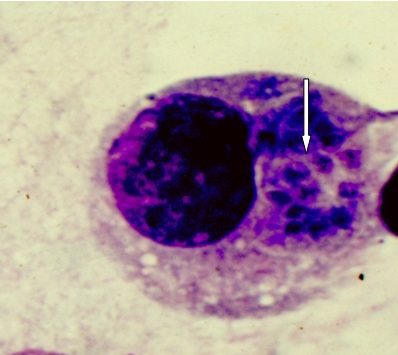
Photo 3. Plate preparation mesentery white mouse at 4 days after intraperitoneal inoculation of a virulent strain of Toxoplasma CDN. Shows the initial phase of the formation of pseudocysts in the cytoplasm of monocytes. Env. Romanovsky - Giemsa.

Env. Romanovsky - Giemsa.

Fig. 1. Besnoitia besnoiti in a skin biopsy from an infected bull (Animal 70) (A) and in material of an infected γ-interferon knockout mouse (K122) (B–G). (A) Numerous cystozoites (<) are released from tissue cysts (*) in bovine skin after squashing using a mortar and pestle. (B) Peritoneal washing of the B. besnoiti infected γ-interferon knockout mouse with numerous extra- and intracellular tachyzoites (<), Giemsa stained. (C–E) Parasites were observed in blood smear of the GKO mouse (Giemsa stained), either in monocytes (C, <), or in neutrophil granulocytes (D, >) or extracellular (E, <) 5 days post-infection. (F) Parasitophorous vacuole containing B. besnoiti tachyzoites arranged as a rosette (<) in a lung section of the infected GKO mouse, H&E staining. (G) Cluster of tachyzoites (>) in a blood vessel of the liver of the GKO mouse, H&E staining.
Photos taken from the German researchers «First in vitro isolation of Besnoitia besnoiti from chronically infected cattle in Germany» ☆
- G. Scharesa, , ,
- W. Bassoa, b, c,
- M. Majzoubd,
- H.C.E. Cortese,
- A. Rostaherf,
- J. Selmairg,
- W. Hermannsd,
- F.J. Conrathsa,
- N.S. Gollnickh
- a Friedrich-Loeffler-Institut, Federal Research Institute for Animal Health, Institute of Epidemiology, Wusterhausen, Germany
- b Laboratorio de Inmunoparasitología, Facultad de Ciencias Veterinarias, Universidad Nacional de La Plata, La Plata, Argentina
- c Consejo Nacional de Investigaciones Científicas y Técnicas (CONICET), Buenos Aires, Argentina
- d Institut of Veterinary Pathology, Ludwig-Maximilians-Universität, Munich, Germany
- e Laboratório de Parasitologica, ICAM, Núcleo da Mitra, Universidade de Évora, Portugal
- f Medizinische Kleintierklinik, Ludwig-Maximilians-Universität, Munich, Germany
- g Inning am Holz, Germany
- h Clinic for Ruminants, Ludwig-Maximilians-Universität, Munich, Germany
http://dx.doi.org/10.1016/j.vetpar.2009.04.033How to Cite or Link Using DOIPermissions & Reprints
Volume 163, Issue 4, 26 August 2009, Pages 315–322
Special Section: EVPC 2008: Veterinary parasitology and climate change
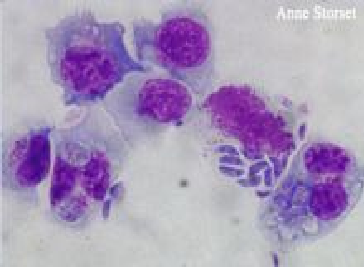
Photo 6. Tachyzoites (trophozoites) Neospora caninum in dogs lymphocytes. Photo is taken from Jackie Barber. Journal Waltham 1998. www.sciencedaily.com/.../11021107.
<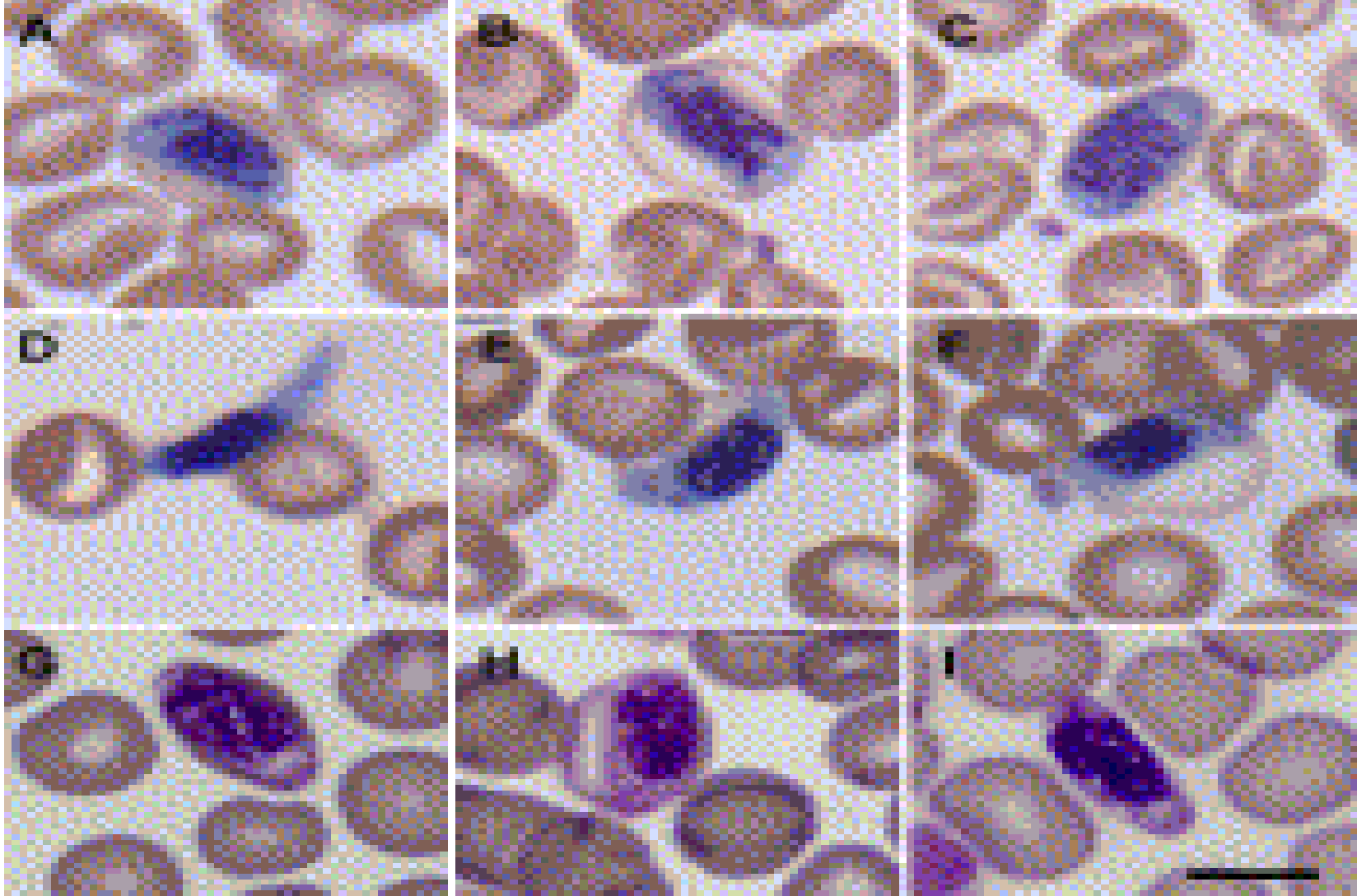
Photo 7. Photo is taken from Ashlie Hartigana, 1, Bing Y. Zhua, 1, Damien P. Higginsa, Paul J. Canfielda, Jan Šlapetaa, published in the journal Veterinary Parasitology Volume 159, Issue 2, 5 February 2009, Pages 105 - 111, made in Australia. The pictures show sarkosporidii in eritrotsinah.
Fig. 1. Intraerythrocytic parasite (Sarcocystidae; Coccidia; Apicomplexa) in thin blood film. Yellow-bellied glider (Petaurus australis) from Queensland (A–F) and New South Wales (G–I). Intraerythrocytic stages are elongate with an oval shape (A, C and H), some displaying a cylindrical outline (B). Centrally located nucleus is coarsely granular. Emerging parasites take elongated form (I), some with one an extended end (D). Before the emergence of these extracellular forms, the parasite starts to slightly elongate at both ends, with the erythrocyte revealing a piriform shape (E–H). The blood films were stained with Quick Dip (Fronine, Australia)
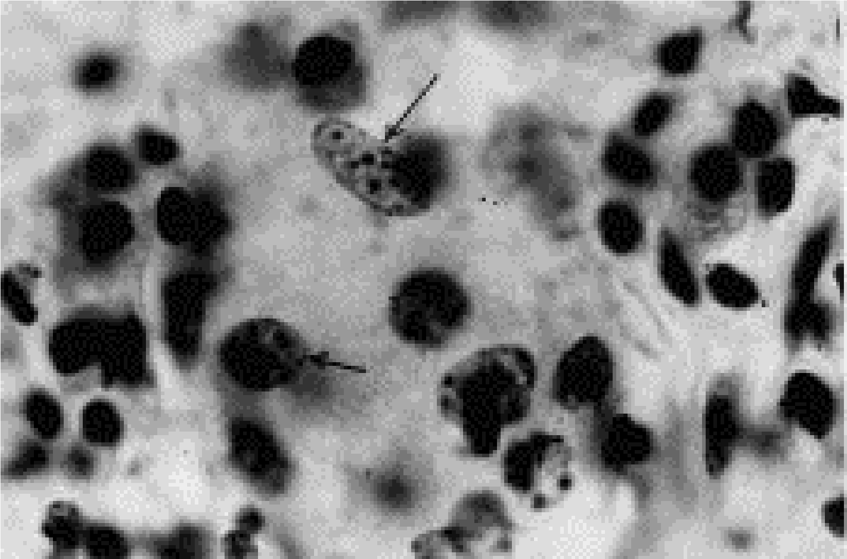
Photo 8. Histological sections of skin goby, subacute phase beznoitioza. See different stages of reproduction beznoity inflammatory cells proliferata - intranuclear reproduction beznoity in gistiotsite (shown by two arrows) and in the cytoplasm of lymphocytes (arrows). In parasites can clearly distinguish between the nucleus and cytoplasm. Okr.Zheleznym hematoxylin and eosin.
For some toksoplazmid characteristic is two independent cycles ramnozheniya - sexual, carried in the body of the definitive hosts and asexual, carried in the body of intermediate hosts. Due to asexual reproduction and development cycle, toksoplazmidy can circulate among wild and domestic animals for a long time, almost endlessly and thus support the existence of species in nature. For such toksoplazmid as Toxoplasma, Neospora, sexual phase of development is not required. And beznoitioza pathogen of cattle (Besnoitia besnoiti) sexual development phase is absent.
Lifecycle toksoplazmid, more similar to the lifecycles gemosporidy and displays the entire evolution of this group of parasites - from free-flagellated plasmodium malaria to humans and beznoity cattle. Therefore, the roots of origin toksoplazmid should look at - does not seem to intestinal protozoa of mammals and of flagellates.
2. Definitive or final boss - it's an animal or a human, in which parasites reproduces sexually, and reach sexual maturity. Toxoplasma is for representatives of all the animals of the cat family.
3. Intermediate host - an organism in which the parasites multiply asexually. Toxoplasma is for all kinds of mammals and birds.
4. Transit or transport host - an organism in which the parasites do not replicate and are stored in the form in which they got to him. Toxoplasma is usually for worms, flies, cockroaches, snails, serving food in small mammals and birds.
5. Obligate host - is host to which the parasite evolutionary and environmentally adapted.
5. Optional boss - is host to which the parasite phylogenetically not adapted, it has poor viability and low fecundity. Example, rodents for Isospora felis and Isospora rivolta. .
6. Monoksennye parasitic protozoa - are parasites who have sexual and asexual reproduction phase are in the same body of the animal (Example, Amery).
7. Polixena parasitic protozoa - are parasites who have sexual and asexual reproduction phase are held in different animal species (Example Toxoplasma gondii and other toksoplazmidy).
2. Toxoplasma gondii - single-celled microscopic parasite that attacks all species of mammals (including humans) and birds. Refers to a type of protozoa, subtype Apicomplexa, has a complex cycle of development and reproduction, both within the host cells and extracellular. It is in the life cycle of Toxoplasma mapped the entire evolution of parasitic protozoa subclass Haemosporina. This is supported by many facts:
First - the existence and propagation of the cells is Toxoplasma - blood plasma, lymph, cerebral white matter, between the fibers of the connective tissue and smooth between the fibers.
Second - propagation of Toxoplasma inside blood leukocytes - neutrophils, lymphocytes and monocytes. There is evidence of breeding birds of Toxoplasma in erythrocytes and erythrocytes of chick embryos in experimental infection (I.G.Galuzo, S.I.Konovalova, 1974, in the book: The life cycle of Toxoplasma, article 84).
Third - Toxoplasma multiplication by direct division of the mother cell to two subsidiaries.
Fourth The presence of flagella microgametes
Fifth Copulation gametes.
Despite the similarity with Toxoplasma gemosporidiyami primarily with Leishmania, Toxoplasma gondii has stringent characteristics distinguishing these parasites from other protozoa, including toksoplazmid.
First - Toxoplasma - it Polixena parasitic protozoa,
adapted to parasitize not only intracellularly in blood cells, hepatocytes, cells of RES and sarcoplasm of muscle fibers, but also extracellularly in - white matter of the brain, blood plasma and lymph, between smooth muscle and connective tissue fibers.
Second - Unlike other parasitic protozoa Toxoplasma at how the body of the definitive and intermediate hosts, there is clearly deterministic stages of asexual reproduction. Asexual reproduction can occur indefinitely, circulating among animals, bypassing the sexual phase.
Pimechanie. All spore-forming parasites, even gemosporidy, asexual reproduction is done is not an infinite number of times, it Toxoplasma can continue indefinitely. Proof - maintaining decades in vitro vegetative forms for Toxoplasma sensitive laboratory animals and in cell culture. To maintain the species in the wild spore usually needed following the asexual stage of sexual development. For Toxoplasma it is not required.
During the multiplication of Toxoplasma in the gut even felines, Toxoplasma individuals of the same type (in the terminology of Dubey and Frenkel, 1972) can reproduce itself, whereas no such ability coccidia. In coccidia schizonts, for example, the first generation can not reproduce themselves such, they should give offspring (merozoites) already belonging to the next generation.
Third. - All Sporozoa initial stage of the development cycle and reproduction is falciform body, often in Toxoplasma - trophozoite, merozoite, and only occasionally bradizoit falciform body.
Fourth. Lack of direct reproduction cycle in the intestine difinitivnogo host - from izosory (oocysts) to izosory (oocysts).
Fifth. The ability of any phase of development and reproduction (except gametocytes and nesporulirovannyh zigotsist) infect animals and humans in any way that enter the body, even intracerebral.
Sixth. Ability to infect izospor Toxoplasma animals and humans both orally and parenterally - subcutaneously, intraperitoneally, intramuscularly, intracerebrally, which is not the case with the so-called "true coccidia."
Seventh. Dualism of development and reproduction in the organism Toxoplasma bradyzoites definitive hosts - davt onset of sexual reproduction phase in the intestinal epithelium and simultaneously abenteric, asexual (trophozoite and cyst phase of reproduction) in other organs and tissues of the same animal.
Eighth. Lack of definitive host sexual reproduction phase when administered orally trophozoites of virulent strains, such as strain RH. While the total of infected animals occurs.
Ninth. Ability to grow and multiply in the body of many species, including humans.
Tenth. Triple mechanism of transmission from donor to recipient - the first by contamination through the cysts and trophozoites izospory that penetrate into the body of animals either orally or parenterally; second, in utero - from mother to fetus through the placenta; third, by predation - predator - prey.
Eleventh. Animals infected not only from individuals of the same species but from different species of mammals and birds.
2.Toksoplazmidozy - in the broadest sense - is the disease caused toksoplazmidami. Our taxonomy - parasites belonging to the order Toxoplasmida. However, in practice, the diagnosis toksoplazmidoz none of the doctors wrote. And rightly so. Although, theoretically, this term has a right to exist, as, for example, the term "Coccidiosis" uniting all diseases caused by parasites belonging to the order Coccidiida. The basis toksoplazmid name (if the diagnosis is written in Russian) takes the generic name is added to the end of the parasite and (Lake). For example, in the basis of the name of the disease caused by Toxoplasma, whichever is the generic name of the parasite-Toxoplasma and added (Lake), resulting in a disease name - toxoplasmosis. The same goes for other diseases caused toksoplazmidami - beznoitioz, neosporoz, gammondioz, frenkelioz, isosporiasis, sarkotsistoz
If you want, for example, when detecting fecal sporocysts sporocysts sarkosporidy emphasize the origin, then write or Sarcocystis bovifelis Sarcocystis bovicanis etc. But this terminology is acceptable in experimental studies, when you know what the animal is taken from tissue cysts. In practice, the detection of sporocysts in the feces of a dog or cat, a doctor is difficult to determine what kind sarkosporidy sporocysts belong to the bovine, ovine, or other types of farm and wild animals. And indeed for the treatment of animals (dogs and cats), this definition is not very important. Therefore, it is wise to ask your physician if sporocysts detected in the feces of an animal, based on the diagnosis of writing, take the generic name of the agent - Sarcocystis and write in prison - SARKOTSISTOZ.
3. Amery or "typical coccidia" - a large group of strictly intracellular protozoan parasites of animals and humans. Amery only infest the intestinal epithelial cells, bile ducts and kidney epithelium. Unlike toksoplazmid have strictly determined ejmery all stages of asexual and sexual development. They reproduce only by schizogony, with the subsequent decay of the schizont on merozoites. Do ejmery, after several generations of asexual necessarily sexual process occurs. (Toxoplasma same can endlessly reproduce asexually, as in the body of intermediate and definitive hosts and maintain a population in nature, bypassing the sexual phase of development). There are only ejmery infective sporulated oocysts contain four sporocysts, each of which is formed by two sporozoites. Contamination can occur only by oral contact with sporulated oocysts in the animal or human body. Amery - it monoksennye parasitic protozoa.
4. Coccidiosis or eymireozy - this disease vyzyvemye Amery or "typical" coccidia - parasites belonging to the order Coccidiida.
Note. In our proposed taxonomy toksoplazmid, parasitic protozoa that form zigotsistah in two disputes with four sporozoites are highlighted in a separate family unit Toxoplasmida. This was due to the fact that such as izospor
I. Felis and I. rivolta, in the body of cats and dogs have been found (Dubey, Frenkel, 1972), along with intestinal stages of asexual reproduction, extra-intestinal stages of parasites (Development, but not breeding!). This cycle of vegetative growth is similar (but not identical!) With those of Toxoplasma and undeniable.
5. Trophozoite or tachyzoite - rapidly proliferating stage toksoplazmid. Tachyzoites can reproduce both intracellularly and extracellularly, as in the cytoplasm of cells and their nuclei. In the trophozoite stage of a rapid rise in the number of parasites in the host organism. Food trophozoites is due to the structure of the hydrolysis infested cell cytoplasm or the trophozoite tissue of the host, if the parasite is extracellular.
6. Homogenised mass - is the cells and tissues of the host organism subjected to hydrolysis by enzymes secreted toksoplazmidami. Ultimately, homogenized represents low molecular weight components are used for further synthesis parasites own nucleic acids, proteins, carbohydrates and other substances necessary for nutrition, parasites and cystic formation of the shell. Gidroliziruya surrounding tissue toksoplazmidy provide themselves with the necessary nutrient material for the development and reproduction (Photo 9 - 12).
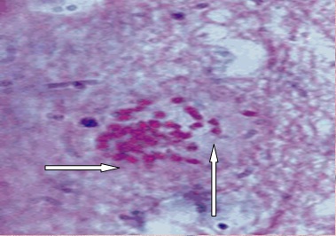
Photo 9. Histological sections of the cerebral cortex of white mice experimentally infected with Toxoplasma slabovirulentnogo strain "LEI". Displaying homogenized tissue (hydrolysis) of the white matter of the brain around the extracellular bradyzoites. Approx. by McManus.
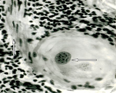
Photo 10. Histological sections of skin goby spontaneously infected Toxoplasma similar parasitic protozoa Besnoitia besnoiti.
Displaying core - pseudocysts smooth muscle cells, in which there is reproduction of parasites. Around pseudocysts visible broad band hydrolyzed surrounding tissue - the smooth muscle fibers and nuclei of the inflammatory infiltrate. Stained with iron hematoxylin and eosin.
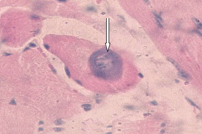
Photo 11. Histological slice right ventricular zebu. Cuba, 1981. Natural infection sarcosporidiosis animal. In the sarcoplasm of the muscle fiber is seen fissile metrotsit around which the visible area homogenized sarkrplazmy painted basophilic. Env. Hematoxylin - eosin.

Photo 12. Histological sections infarction zebu. The photo shows two cysts formed in the sarcoplasm of muscle fibers Sarcocystis sp. The left in the photo is visible to a small colony of parasites with a wide shell and two membranes - internal dividers separates parasites from a wide basophilic layer and outer layer separates from a basophilic modified (hydrolysed) sarcoplasmic muscle fibers. Hydrolysed sarcoplasm around the colony of parasites eosin stained intensely. Right, shows a colony of parasites, which can be seen a large number of parasites dividers basophilic staining. Around the colony, basophilic cladding layer cysts stored and homogenized sarcoplasm of the muscle fiber is squeezed and is a small eosinophilic rim. Env. iron hematoxylin and eosin.
Honors homogenized sarcoplasmic muscle fibers located around metrotsitov Sarcocystis, homogenized from surrounding tissue formed around the nuclear pseudocysts Besnoitia besnoiti is homogenized sarcoplasm that gives a positive reaction to sharply Brachet RNA. While homogenized mass around beznoitioznyh pseudocysts, although it contains RNA, but still it is dominated by neutral mucopolysaccharides (Photo 13). It should be noted that bradyzoites example, Toxoplasma and beznoity contain more amylopectin than RNA, whereas both in the cytoplasm bradyzoites sarkosporidy found only traces of amylopectin. Whereas RNA fills almost the entire cytoplasm of parasites.

7. Parasitophorous vacuole - a zone of hydrolysis cytoplasm infested cells or tissue surrounding the parasite, where concentrated low molecular building blocks for further synthesis of nutrients necessary for the parasite (Photo 14 - 15).
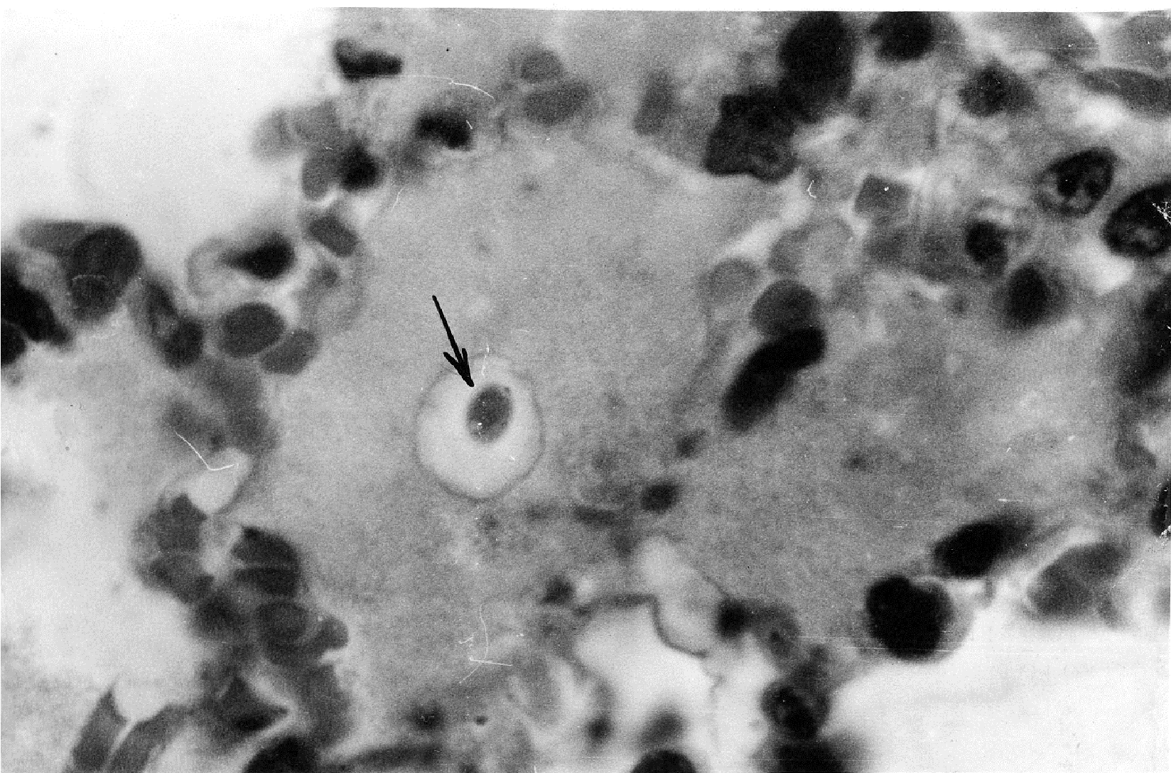
Photo 14. Histological sections of lung puppy who died on the 15th day of acute experimental toxoplasmosis. In serous exudate visible Toxoplasma (tachyzoite) with a well defined parasitophorous vacuole. Env. Hematoxylin - eosin.

Photo 15. On Electron photo shows parasitophorous vacuole around T. gondii tachyzoites located in the cytoplasm of peritoneal exudate cells (cells which authors do not specify). Photo is taken from J. P. Dubey,1,* D. S. Lindsay,2 and C. A. Speer3« Structures of Toxoplasma gondii Tachyzoites, Bradyzoites, and Sporozoites and Biology and Development of Tissue Cysts»†
Transmission electron micrograph of a tachyzoite of the VEG strain of T. gondii in a mouse peritoneal exudate cell. Am, amylopectin granule; Co, conoid; Dg, electron-dense granule; Go, Golgi complex; Mn, microneme; No, nucleolus, Nu, nucleus; Pv, parasitophorous (more ...)
8. Colonial parasitophorous vacuole - this area of the hydrolysis of extracellular tissue of a host organism, formed by hydrolysis around the colony of parasites and serving nutritious substrate for extracellular free bradyzoites, as well as parasites are located inside the nuclei and cysts (Photo 16).
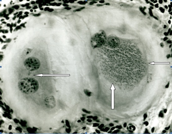
Photo 16. Histological sections of the skin 11 month goby subacute stage spontaneous beznoitioza. Shows two simultaneously forming tissue cysts B.besnoiti hydrolyzed in the surrounding tissue. In one of them, on the left in the photo, you can see three nuclear pseudocysts. In another - three nuclear pseudocysts and a large colony of bradyzoites in a homogeneous mass. Around the colony already visible membrane, which, on the one hand, holds the colony is compact and is bordered by the nutrient mass - the emerging colonial parasitophorous vacuole. Stained with iron hematoxylin and eosin.
In cysts with thick shells, colonial parasitophorous vacuole is formed in the middle layer of the shell. And, - apparently until this layer there is not degenerating cyst. After thinning of this layer is observed in the cyst degeneration tsistozoitov and such cysts are attacked macro - and macrophages, and they die, and in their place are formed granulomas (Photo 17 - 21)
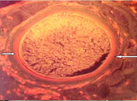
Photo 17. Histological sections of skin goby subacute beznoitioza. Preparation treated with acridine orange. Inner layer (colonial parasitophorous vacuole) fluoresce orange, indicating that the content in it large amounts of RNA.
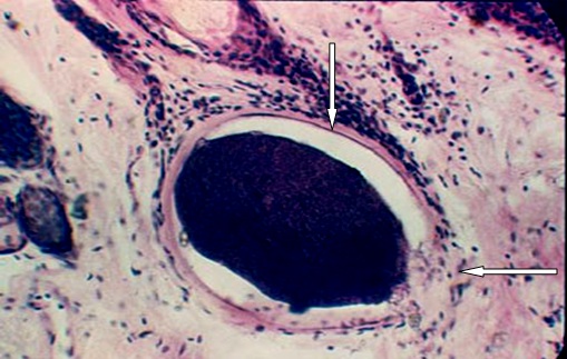
Photo 18. Histological sections of the dermis cow beznoitiozom chronic sick. Displaying tsistozoitov dystrophy, destruction shell cysts and infiltration of inflammatory cells into the cysts. The photographs show clearly the inner envelope membrane cysts that holds the lost colony of parasites. Colonial parasitophorous vacuole is represented as a thin rim of basophilic. Env. Hematoxylin - eosin.
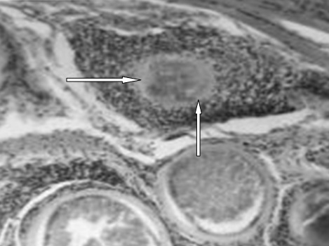
Photo 19. Histological sections of the dermis cow beznoitioza chronic course. Along with well-formed cysts seen perishing cyst surrounded by a large number of cells of the host organism. Env. Hematoxylin - eosin.
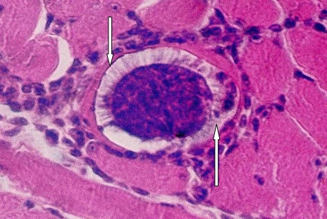
Photo 20. Histological sections of skeletal muscle zebu breed buffalo died from "sudden death" in the province of the Republic of Cuba Bayamo. Displaying dystrophy thick shell cysts sarkosporidy under natural infection and penetration into the cavity of the cyst macrophages. Env. Hematoxylin - eosin.
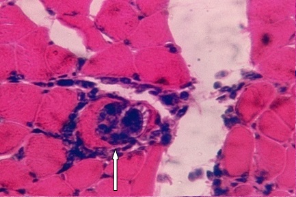
Photo 21. Histological sections of skeletal muscle zebu breed buffalo died from "sudden death." Shows a thick-walled cysts sarkosporidy death and its replacement cells proliferata. Env. Hematoxylin - eosin.
Photo 22. Photos taken from a textbook on pathologic anatomy of farm animals and Niberle Corsa, 1980. The figure shows the death tsistozoitov sarkosporidy penetration and macro - and macrophages into the cysts.
In Toxoplasma cysts and tokostennyh sarkosporidy death cysts on - Videmo, other factors associated with disturbed nutrition in large colonies of Toxoplasma cysts, namely thinning and disappearance pretsistnogo homogeneous layer (colonial parasitophorous vacuoles) (23-25).

Photo 23. Histological sections of skeletal muscle zebu breed buffalo died from "sudden death" in the province of the Republic of Cuba Bayamo. The photo shows a thin-walled cyst sarkosporidy. Places clearly visible thinning pretsistnogo homogeneous layer (colonial parasitophorous vacuoles) and penetration in areas of thinning pretsistnogo layer phagocytes into cysts. Env. Hematoxylin - eosin.
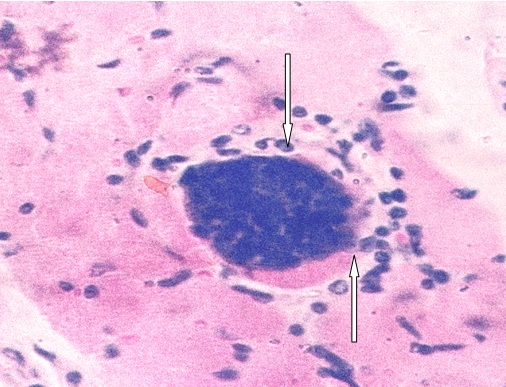
Photo 24. Histological sections of skeletal muscle Zebu bull calf died from "sudden death" in the province of the Republic of Cuba Bayamo. Shows a thin-walled cyst sarkosporidy perishing and ingress macro cysts, and macrophages.
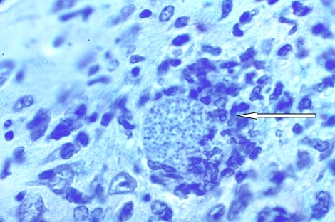
Photo 25. Gistologicheskay brain slice white mouse experimentally infected with a strain of Toxoplasma slabovirulentnym «LEI». Shows the moment of death and phagocytosis cysts. Penetration of micro - and macrophages is observed in a plot of extinction pretsistnogo nutritive layer. Stained with iron hematoxylin.
9. Toksoplazmid Colony - a group of extracellular breeding and developing bradyzoites forming a cyst (Photo 26).
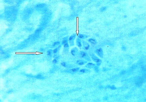
Photo 26. Smear - imprint brain white mouse infected slabovirulentnym strain of Toxoplasma «LEI» and killed 10 days after infection. Shows a small, extracellular Toxoplasma colony in the white matter of the brain stem. Shell around the colony of parasites still missing. Stained with iron hematoxylin with blue tint metelenovym.
Tissue cyst toksoplazmid - a spherical or oblong - oval extracellular colony slowly agamogenetic parasites surrounded clearly contoured shell. Tissue cyst is one of the phases of reproduction and development of parasites. Tissue cysts, for example, Toxoplasma formed not only in the body of animals and humans, but also in cell culture, where there are no antibodies (Gustavson at all., 1954; Hogan at all., 1960, 1961; Beverley, 1969 Galouzeau, Konovalov , 1974; Baron S.1996) .. So, to say that tissue cyst is formed as a result of the defense mechanisms of the host organism for no reason. Cyst - a "Kokan" or "house" built by the parasite by the host material. Granulation tissue of a host organism with no does not participate (Photo 27 -34).
|
|
|
|
|
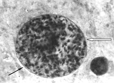
Photo 27. Smear - imprint brain white mouse. Toxoplasma cysts shows with well contoured shell. Inside the cysts seen many bradyzoites. No granulation tissue around the cyst is not visible. Stained with iron hematoxylin and eosin.

Фото 28. A.Tissue cyst freed from mouse brain. Note a thin (arrow) cyst wall enclosing hundreds of bradyzoites. Unstrained. Bar = 20 μm.
B.Two tissue cysts (arrows) in section of brain. Hematoxylin and eosin stain. Bar = 20 μm.
C.Transmission electron micrograph of a small tissue cyst in cell culture. Note thin cyst wall (arrow) enclosing 6 bradyzoites (arrowheads). Bar = 1.0 μm. (Courtesy of Dr. D.S. Lindsay, Auburn University, Auburn, AL.)
The pictures show extracellular Toxoplasma cysts in the brain experimentally infected white mouse and the intracellular formation of cysts in cell culture.
Photo taken from the article Baron s. (1996).
Medical Microbiology. 4th edition.
Baron S, editor.
Galveston (TX): University of Texas Medical Branch at Galveston; 1996.
Copyright © 1996, The University of Texas Medical Branch at Galveston.NCBI Bookshelf. A service of the National Library of Medicine, National Institutes of Health.

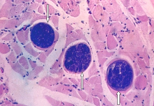
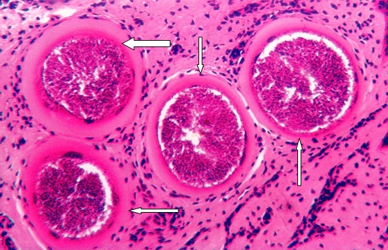
Photo 31. Histological sections of the dermis in chronic beznoitioze cows. Showing a well-formed extracellular cysts B.besnoiti with multiple bradyzoites. Around cysts visible only homogeneous eosinophilic mass. Connective tissue no part in this does not take apart the fabric hydrolyzed. Env. Hematoxylin - eosin.
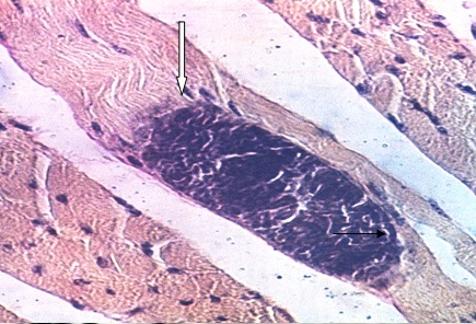
Photo 32. Histological sections of skeletal muscle 14 month bull calf breed zebu fallen from "sudden death." Shows a longitudinal section through the atrophied muscles of the beam emerging from it sarkosporidy extracellular cysts. Env. Iron hematoxylin and eosin.

Photo 33. Histological slice right ventricular zebu. Showing a well-formed cysts sarkosporidy in muscle fibers (arrows) and nerve fibers (shown by two arrows). Env. Hematoxylin - eosin.

Photo 34. Extracellular, thick-walled cyst in the brain is undeniable rottvelera biennium. Photo is taken from Jackie Barber. Journal Waltham 1998.
www.sciencedaily.com/.../11021107.
8. Bradyzoites - it slowly propagate extracellular parasites that form tissue cysts. Morphology, they are of two types. Some nuclei with a central location, with other nuclei located a blunt edge, the last look reminiscent parashutika. Theoretically and practically, you can call the bradyzoites and parasites inside the cysts, as the parasites continue to multiply slowly and after the formation of a shell around the colony of parasites. However, it is more convenient and easier to understand, parasites in cysts formed tsistozoitami call, as it is accepted by most researchers.
Thus, when the light-optical study long been observed that tissue cysts of Toxoplasma contain parasites at different stages of development (Photo 35). Like other toksoplazmid, training occurs in cysts of parasites in sexual development and reproduction. Therefore depends on the maturity of the cyst and prepatentny patent period in the intestines of cats.
Researchers studying Toxoplasma cysts and their relationship with the host, never saw that bradyzoites cause necrosis of the surrounding tissue. Bradyzoites only cause gomogenezatsiyu hydrolysis or rather the surrounding cells and tissues, but not necrosis. Hydrolyzing the surrounding tissue bradyzoites thereby prepare the environment, for the subsequent synthesis of her nutrients needed parasites are in the colony. Bradyzoites differ from tachyzoites not only antigenic structure and rates of reproduction, but also metabolism, tropism and pathogenicity. For example, tissue necrosis may cause only tachyzoites. So when some authors write that the necrosis part bradyzoites, it only shows that the writer does not understand the doctor, differs from tachyzoite bradyzoites.
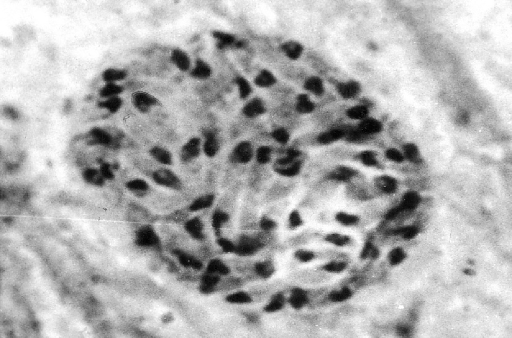
Photo 35. Smear - imprint brain white mouse experimentally infected slabovirulentnym strain of Toxoplasma «LEI». Shows a group of extracellular bradyzoites in the formation of cysts. Bradyzoites seen having different morphological structure and are at different stages of division. Shell around the colony is still missing. Stained with iron hematoxylin.
In contrast to tachyzoites, as reproduction, and colony formation in the cytoplasm of certain types toksoplazmid gradual accumulation amilipektina that changes not only by their morphology, but the biological purpose. For example, Besnoitia besnoiti located on the periphery of the colony, accumulated in the cytoplasm of bradyzoites amylopectin, is to build the inner lining of cystic wall (Photo 36)

Photo 36. Histological sections of skin goby subacute beznoitioza. Shows a portion of the beginning of the formation of the inner membrane of cystic shell Besnoitia besnoiti. Good breakup can be seen on the periphery of the colony bradyzoites and release granules while amylopectin. Staining Mac - Manus.
It is possible that and Toxoplasma cysts in education shell participates as hydrolyzed weave white matter of the brain, and amylopectin accumulated bradyzoites. This is indicated by a positive reaction shell cysts on Mac - Manus in the initial stages of its formation.

Photo 37. Gistosrez brain white mouse, 15 days after intramuscular infection slabovirulentnym strain LEI. Around the colony Toxoplasma clearly visible thin, Chic - positive shell. Staining Mac - Manus.
In more mature cysts of Toxoplasma cysts shell changes its chemical composition and color on Mac - Manus longer gives a positive reaction to amylopectin and stained basophilic (Photo 38).
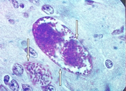
Photo 38. Histological sections of the cerebral cortex of a white mouse for 20 days after intramuscular infection slabovirulentnym strain LEI. Shows well formed extracellular cyst, with a clearly visible fine basophilic shell. Nearby, in the white matter, seen a small colony of extracellular Toxoplasma. Cytoplasmic bradyzoites gives intense reaction to amylopectin. Env. For Mac - Manus.
Stained histological Mallory, using phosphotungstic hematoxylin shows that the formation of cysts shell participates and hydrolysed homogeneous (mucous) weight. Mallory method gives shell cysts characteristic of the hydrolysis reaction of the tissue and is painted in dark - blue. It should pocherknut that gistosrezov when stained by this method is colored in blue and collagen. However, the appearance of collagen around the colony Toxoplasma no reason, since no cysts formed around the cell reaction is not observed. (Collagen is known to be formed by fibroblasts, and around their colonies no Toxoplasma approx. Authors). Interestingly, karyenchima bradyzoites stained Mallory in light - pink, bright orange nucleoli - red and cytoplasm - in light - blue (Photo 39).
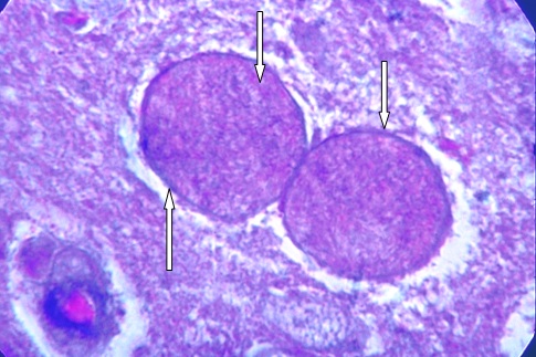
Photo 39. Histological sections of the brain of a white mouse on day 20 after intramuscular infection slabovirulentnym strain LEI. Shows the response of shells and Toxoplasma cysts themselves tsistozoitov when stained slice method with Mallory phosphotungstic hematoxylin. Clearly visible dark - blue color shells cysts, light - blue cytoplasm and bright orange color tsistozoitov nucleoli.
6. Tsistozoity - it slowly propagate parasites inside the cysts. Term tsistozoit indicates the location of the parasite and can be applied when it is necessary to emphasize that bradizoit is well-formed cyst. Tsistozoity and bradyzoites differ from tachyzoites or trophozoites, as rates of reproduction, and content in their cytoplasm amylopectin. If cytoplasmic RNA predominates Toxoplasma trophozoites, the bradyzoites amylopectin (Photos 40 - 41). In recent years found that they differ in antigenic structure.
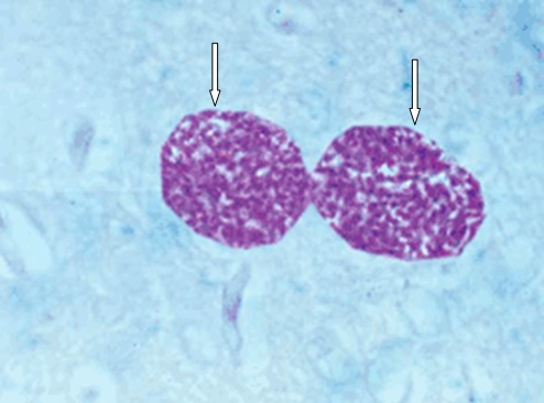
Photo 40. Histological sections of the brain of white mice infected with Toxoplasma slabovirulentnogo strain of Toxoplasma. Shows two well-formed cysts, cysts shell painted basophilic, while sharply tsistozoity give a positive reaction to amylopectin. Staining Mac - Manus.
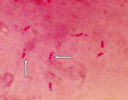
Photo 41. Smear-mark white mouse brain infected slabovirulentnym strain of Toxoplasma. Displaying uneven distribution of amylopectin in the cytoplasm tsistozoitov. In some tsistozoitov amylopectin located predominantly in the rear part of the body, in others it is seen as the rear and in front of the parasite cells. Staining Mac - Manus.
8. Toksoplazmid shell cysts - is a complex organized extracellular parasites colony structure, providing protection from aggressive tsistozoitov and promotes pitataniyu mnogotychyachnoy colony parasites. Shell cysts in toksoplazmid formed in three ways:
First - is the transformation of the parent shell in a shell parasite cysts in endodiogenii, endopoligenii and schizogonic; Second - is the formation of the shell of the host tissues hydrolyzed amylopectin and parasites themselves; Third - often observed in Besnoitia besnoiti - is the transformation of the nuclear envelope (nuclear pseudocysts) in shell cysts.
It is especially necessary to emphasize that the granulation tissue to shells cysts toksoplazmid has no relation. Therefore shell cysts toksoplazmid better not call a capsule. Since the term "capsule" in the body of animals imply the formation of connective tissue origin. For example, the capsule around a small foreign body or connective tissue capsule around the larvae of helminths (chalicosis) or connective tissue capsule around the primary tuberculous focus, etc.
9. Pseudocysts - this is one of the phases of development of intracellular toksoplazmid, often in cells RES. Pseudocysts - is not just a cell infested with parasites. This Intracytoplasmic or intranuclear colony of parasites, formed from a single mother cell by schizogonic, endodiogenii or poliendogenii. Pseudocysts formed only in a parasitophorous vacuole in the formation of which occupies an important place sheath cytoplasm or nucleus infested cells (Photo 42 - 43). This is the last stage of asexual, intracellular multiplication toksoplazmid observed in subacute and chronic phase of the disease animals. This stage is followed by a phase of extracellular toksoplazmid reproduction and development, the formation of tissue cysts reproduction. Typically, pseudocysts do not turn into cysts. As nakoplaniya parasites in the cytoplasm, the shell infested cells destroyed and the parasites leave the dead cells. In pseudocysts parasites accumulate nutrients for later reproduction in extracellular brain, muscles and other organs in which they form tissue cysts reproduction.
In some cases, for example in Besnoitia besnoiti, nuclear pseudocysts turns into tissue cyst. Thus there is a complete atrophy karyoplasm infested cells and cytoplasm. Shell same nucleus is transformed into a shell cysts. This shell has been a different structure and functionality, namely, it provides, on the one hand protect the colony of parasites from the pathogenic effects of the host organism, on the other - facilitates the processes of assimilation and dissimilation thousandth colony of parasites inside the cysts.

Photo 42. Histological sections of liver puppy experimentally infected with a virulent strain of Toxoplasma CDN. Shows a typical pseudocysts in Kupffer cells. Env. Iron hematoxylin and eosin.

Photo 43. Photo taken from work Alexandre Paulino Loretti, and Severo Sales Barrosb «Hemorrhagic disease in dogs infected with an unclassified intraendothelial piroplasm in southern Brazil», published in 2005 in Brazil.
Shows a typical pseudocysts in kidney endothelial cell (cell RES) in experimental dogs piroplasmosis.
(Bone marrow, dog, experimental case. Smear. Many zoites are found in the cytoplasm of an endothelial cell. Panoptic. Bar = 4.5 μm)
10. Infested cell - In acute during toksopdazmoza in histological sections, smears - imprints of the bodies in the blood smears in the mesothelial cells of the peritoneum and pleura can find a lot of infested cells. Often multiple cells simultaneously invading trophozoites, which give rise to both binary and multiple fission (schizogony). The result of this invasion is cell death and education mikronekrozov with numerous parasites around the necrotic tissue. Infested cell, and especially lots of Toxoplasma Toxoplasma never turns into pseudocysts, it collapses and parasites come into the extracellular space and give rise to a new generation of parasites. Often, together with the collapse of infested cells die and parasites themselves. If the invasion and destruction of the mesothelial cells that are exposed in the abdominal exudate out parasites at different stages of development and reproduction (Photo 44 - 45).

Photo 44. Plate preparation mesentery white mouse. Displaying mesothelial cells with simultaneous multiple infestations. Toxoplasma are at different stages of binary and multiple division. Around each group of parasites has its parasitophorous vacuole. Colouring Romanovsky - Giemsa.
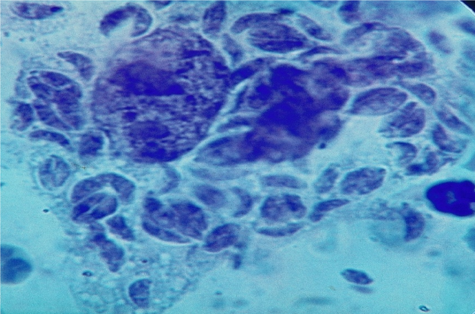
Photo 45. Plate preparation mesentery fallen white mouse on day 6 after infection. Displaying mesothelial cells simultaneously invade a variety of Toxoplasma. Infested cells vacuolated cytoplasm, sometimes sheath cells destroyed and parasites freely leave the cell. Okr.zheleznym hematoxylin and eosin, with methylene blue tint.
10. Merozoite - a stage of the life cycle of parasites formed as a result schizogonic.
11. Zigotsista - it nesporulirovannaya zygote. Zigotsisty, released into the environment, along with the feces after sexual reproduction phase toksoplazmid in the epithelium of the small intestine, canines and felines.

Photo 46. Zigotsisty (I.felis), in a five-month kitten feces 4 hours after their release into the environment. Most of zigotsist not sporulated. Natural infection.
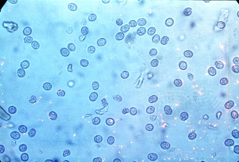
Photo 47. Displaying more quantity zigotsist Toxoplasma in feces experimentally infected kitten 3 minutes after their release from the intestine. Such zigotsisty not dangerous to other animals and humans.
Zigotsistami catch can neither man nor animal, they are not invasive (non-infectious) education. They are no sporozoites kotorve should be formed by sporulation in the external environment. Zigotsista term for Toxoplasma introduced academician I.G.Galuzo to emphasize the difference between Toxoplasma and coccidia. However, this term may legitimately be applied to other generators izospory parasites. Zigotsisty in the environment, with a good supply of oxygen sporulate and become invasive (contagious).
13.Izospora - it sporulated zigotsista. As a result of sporulation in izospore formed two disputes with four sporozoites each. Izospory formed in the external environment, the presence of oxygen in 15 - 20 minutes after hitting them in the external environment! Some works are evidence that izospory formed after 5 - 6 hours - it may due to poor aeoratsiey zigotsist. Under normal household conditions and natural sporulation begins after 15 - 20 minutes. Given this rate of sporulation zigotsist Toxoplasma, for their own safety, cat feces should be removed as quickly as possible.
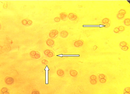
Photo 48. Showing sporulated izospory ((I.felis) and T. gondii)
isolated from feces 2-month kitten with mixed infestations.
11.Amilopektin - One of the main starch polysaccharide, consisting of the branched molecular chains glyuklzy. He serves as the main nutrient and structural material for toksoplazmid, especially in a phase of formation of tissue cysts and pseudocysts breeding. Amylopectin role in different stages of development toksoplazmid uncertain. If Toxoplasma and beznoity, as their intracellular multiplication, amylopectin accumulates in the cytoplasm of the parasite and its maximum stated in bradyzoites, then sarkosporidy forming a cyst or cysts are missing amylopectin.
On the situation in the system simple toksoplazmid.
Systematics of the simplest is to some extent artificial construct, as a basis for its various features are taken seem important at some stage of our knowledge about animals. And she (taxonomy) changes, as there are more and more data on the structure and life cycles of parasitic protozoa. So, initially, based on the classification of elementary bodies were taken to the movement. And all just split into 4 classes. First class: Sarcodina or Rhizopoda; second class: Sporozoa; third grade: Mastigofora and fourth - Infusoria.
Then (Doflein, 1902) as the basis of classification of parasitic protozoa put "organs of fertilization and reproduction" and the first three classes combined into one group, subtype Plazmodroma, and took it to the Infusoria subtype Ciliophora. However, such an approach could not, no doubt, to unite all the parasitic protozoa in the system was created. With this approach, the main controversy arose when determining gemosporidy and in particular members of the group Plasmodium. If we take as a basis for "sexual characters" that malaria parasites would have to join the group of Coccidia, although the similarity of the two groups is only in the fact that malaria parasites have a sexual phase, culminating in the formation of spores in the mosquito. Talk about full identity can not be out of question, because the "true" coccidia live in the epithelial cells of humans and animals. Shizogoiya, education gametocytes and zygote formation in coccidia occur in the same vertebrate host and sporulation in the external environment. In Plasmodium falciparum schizogony and gametogony occurs in blood cells - white blood cells, red blood cells and cells of the RES and hepatocytes vertebrate host, and the fertilization and sporogony in the other (definitive) host, which is a carrier - insect (mosquito). "Sexual approach" could not make clear to other groups of blood parasites such as piroplazmy, Tayler and members of the genus Leucocytozoon. According to the "sexual" classification beznoitii, toxoplasma, sarkosporidii, Tayler, piroplazmy were placed as parasites of uncertain about the nature of class Sporozoa. Then the basis of systematics of parasitic protozoa began to take not just one, which is a definite sign, and the entire life cycle of the parasite, including its morphology. As a result of this approach gemogregariny, gemosporidii, toksoplazmidy, plasmodium piroplazmy and were in the same suborder (suborder approx.'s) Haemosporidia order Coccidiomorpha (N.Valiyev, 1930).
This taxonomy class Sporozoa held until 1967 the last century. In 1967, EM Heysen modified sistimatiku parasitic protozoa and singled in two subclasses of class Sporozoa - Gregarinina subclass and subclass Coccidiomorpha three companies - Protococcidiida, Adeleida and Coccidiida. Detachment Coccidiida identified two suborder - Eimeriidea and Haemosporidiidea, which includes all parasitic protozoa class Sporozoa.
According to this classification of the subclass Gregarinina became known gregarine and the subclass Coccidiomorpha coccidia. However, in the narrow sense, typical or classic coccidia are usually only representatives of the family Eimeriidae. EM Kheysin not included in the class toksoplazmid Sporozoa, and supported the proposal of E. Biocca (1956) on the allocation of a separate class toksoplazmid Toxoplasmatea inside subtype Plasmodroma.
This taxonomy of scientists held until 1969. In the early 70 - ies in Toxoplasma was discovered sexual phase of development (Hutchison et al. 1969, 1970; Frencel et al.1970; Overdulv, 1970) and Sarcocystis (Rommel et al.1972; Rommel and Heydorn.1972) . It was found that the sexual phase of development in Toxoplasma and sarkosporidy performed like Amery. However, it is not identical to Amery. Despite this, some researchers again as they did 70 years ago, was taken as the basis of classification one characteristic - sexual and as a result of this approach, all toksoplazmid, including Besnoitia besnoiti (crowd) is placed in the subfamily in the family Isosporinae Eimeriidae (Beyer et al .1978). However, in 1970, ND Levine (Levaine) in the preparation of the classification system of parasitic protozoa, based his ultra-structure of parasitic protozoa Sporozoa class and combined them into a subtype Apicomplexa. The system ND Levine beznoitii cattle (B. besnoiti) placed as a separate genus in the family Sarcocysidae.
With the inclusion of Toxoplasma and all in the suborder toksoplazmid Eimeriidea family Eimeriidae categorically disagreed Academician I.G.Galuzo. He in 1970, after the discovery of the sexual phase of development in Toxoplasma (T.gondii) and sarkosporidy (S.fusiformis) toksoplazmid identified as an independent unit in the classroom Sporozoa. On the selection of an independent toksoplazmid taxonomic group, at a high level, and fought Professor DN Zasukhin.
Based on the data of the time (late 60's and early 70s), IG Galouzeau summed so grand total research of that time in his book "The life cycle of Toxoplasma" which was published in 1974: "First of all, it is necessary to withdraw the question, which is often put before us: Toxoplasma coccidia or not? And tantamount him izospora Toxoplasma or not? Apparently, neither one nor the other. When all toksoplazmopodobnye organisms (known and unknown) will find their "buddy" of Eimeriidae, when there will be many more ambiguities resolved, in particular the mechanism of proliferation in coccidia when they, with a focus on research on the life cycle of Toxoplasma, revised mechanisms of coccidia, then finally resolved the issue of taxonomists exciting place toksoplazmid, as well as coccidia, in the simplest system. Meanwhile, now it is obvious that as the nature of the sexual and asexual phases of development of Toxoplasma, which has significant differences with the known development plan coccidia, and the behavior of those and others in nature, these two groups of organisms are in considerable taxonomic distance from each other " .
Unfortunately, this book was the last work in the life of IG Galouzeau and given to the conclusion of the chapter "On the unity of nature and Toxoplasma izospor" became, as it were, the main focus will and toksoplazmid study for his students.
Systematics of parasitic protozoa and in particular the proposed toksoplazmid I.G.Galuzo In 1974, in an abbreviated form is as follows:
Class Sporozoa Leuckart, 1879
- Division Coccidiomorpha, Dof llein, 1901
- Order Coccidiida, Labbe, 1899
- Order Toxoplasmida, Biocca, 1957
- Family Toxoplasmidae, Dubey Frenkel, 1970
# Genus Toxoplasma
# Genus Sarcocystis
# Rhode Besnoitia
# Rhode Frenkeiia,
# Genus Isospora
# Genus Isospora,
Since the proposed academician I.G.Galuzo classification has been more than forty years. To date, fully studied lifecycle T.gondii, B.besnoiti, Sarcocyctis, Atoxoplasma, I. felis and I. rivolta. Open sexual and asexual phases of development and reproduction in Neosporum caninum. It appeared that the life cycle undeniable practically identical to those of the c T.gondii. Circle of animals being hit is undeniable about the same as that of Toxoplasma. The only difference is that the definitive host for Toxoplasma are all members of the family Felidae, and for the undeniable (N. caninum), canines - Canidae. Until today, it is not known whether the undeniable hit man. An important discovery was the discovery for Protozoologists have I. felis and I. rivolta extraintestinal stages of development in the body of intermediate hosts that end formation dermozoitov (Dubey, Frenkel, 1972). Dermozoity can give rise, in the intestine difinitivnogo host sexual process culminates in the formation nesporulirovannyh zigotsist (oocysts). This discovery brings the genus Isospora with toksoplazmidami and can not be considered more than I. felis and I. rivolta, as monoksennyh parasites. Discovering I. felis and I. rivolta extraintestinal stages of development gives the right to include these parasites in the squad Toxoplasmida, highlighting them in a separate family.
Obtained in recent years, new data on toksoplazmid once again confirm that this group of parasitic protozoa is markedly different from the so-called "classical or true coccidia," as on endogenous reproduction and development, and on their circulation in nature. And new data give reason to rethink the notion of the place not only beznoity cattle in the simplest system (in which there is no sexual phase of development), but Toxoplasma undeniable, sarkosporidy and other parasitic protozoa. It seems justified to keep the system simple detachment Toxoplasmida. And in memory of the prominent Soviet Protozoologists twentieth century - Dmitry Nikolayevich Zasukhina and Hilarion Grigorev Galouzeau who made a great contribution to the history of protozoology, the founders of the first two centers to study the problem of toxoplasmosis in the USSR squad called their names. A view Besnoitia besnoitii be named Professor BP Vsevolodova, first described these parasites in the USSR.
In view of the above stated, we toksoplazmid system seems to date as follows:
Type Protosoa
Subtype Apicomplexa Levine, 1973
Class Sporozoa Leuckart, 1879
Subclass Coccidiomorpha Dof llein, 1901
Detachment Coccidiida Labbe, 1899
Family Eimeriidae Minchin, 1903
Subclass Haemosporina Danilewski, 1885
Detachment Toxoplasmida Biocca, 1957, Zasuhin and Galuso, 1970
Family Neosporidae Bjerkas, Mohn, Presthus, 1984
Genus Toxoplasma Hatchison, 1969, Dubey, Frenkel, 1970
- Type of T. gondii, Nicolle and Manceaux, Splendore, 1908
- Genus Neospora Dubeu, 1988
- View of N. caninum
Rhode Besnoitia Marotel, 1912
- Type B. besnoitii Marotel, 1912, Vsevolodov, 1959
- View B.jellisoni.
- View B.darlingi
- View B.tarandi
- View B.wallcei
GW Sarcocystidae Ray Lankester 1882
Genus Sarcocystis Rommel, Heydorn, 1972
- View S.fusiformis
- View S.tenella
- Type S. mischeriana etc.
Rhode Frenkelia Biocca, 1968
- View F.frenkeli Rommel, Krampitz, 1975, etc
Family Isosporidae Schneider 1881
Genus Isospora Wenyony 1923.
- Type I. felis
- Type I. rivolta etc.
Detachment Piroplasmida Wenyon, 1926
Detachment Haemosporida Levine, 1961
Detachment Adeleida Leger, 1911
The main signs of Division Haemosporina
- Parasites with a distinct apical complex;
- Parasites white blood count cells of the reticulo - endothelial system and red blood cells;
- Parasites with facultative and obligate geteroksennostyu;
- Broad host specificity of parasites. (The parasite is able to multiply in the body for at least two species of animals - mammals, birds, human, blood-sucking insects and mites);
- Parasites, similar in morphology and life cycles, but who may lack the sexual process and sporulation;
- Parasites that can multiply in the cytoplasm of cells invaded and their nuclei;
- Parasites in the life cycle which alternates phase of intracellular and extracellular growth and reproduction;
- Parasites, sexual development and reproduction phase which is not required, although it is present in their life cycle;
- Development of the zygote goes through sporulation to form 8 or more sporozoites in each zigotsiste (oocyst) or zygote transformation in mobile ookinetu;
- Sporogonia occurs within the host organism and in the external delusions and ends with the formation of sporozoites;
- Sexual reproduction and development phase can take place, as in mammals and birds, and in the body of blood-sucking insects and mites;
- Mandatory (gemosporidii, sarkosporidii) or optional change of the definitive host (Toxoplasma, beznoitii undeniable, izospory, atoksoplazmy, etc.);
The main signs of detachment Toxoplasmida
- Mnogohozyaenny parasite with a complex life cycle and greatly hypertrophied asexual phase of development and reproduction
- The parasite life cycle which includes the reproduction and development in the body of the definitive, intermediate host and the external environment.
- The parasite for which the sexual process may not be required;
- The parasite, which at one stage of endogenous reproduction, colonial development phase has a "specialization" of parasites inside the colony;
- The proliferation of the parasite in the host runs continuously. It may occur, moving from one host to another, without the sexual stage of development and sporogony;
- Parasite, asexual reproduction and development cycle which, in a host organism, comprising the trophozoite phase (proliferation phase) pseudocysts (preparation phase to parasitic formation of tissue cysts reproduction) and the phase of tissue cysts reproduction (slow reproduction phase and maintaining the parasites in the host organism and preparation for their further circulation in nature);
- Parasite in phase trophozoite (proliferation) able to reproduce both intracellularly and extracellularly.
Parasite with intracellular multiplication able to form around him parasitophorous vacuole (feed zone).
- The parasite, in a phase of formation of tissue cysts - is extracellular, colonial organisms forming colonies around parasites nutritional zone (colonial parasitophorous vacuole) - cysts in the wall of the shell or in pritsistnom layer;
- The parasite, which tissue cyst is formed by a complex interaction of biological colony of parasites with the host. Shell form cysts parasites themselves due to "building material" host organism and are, in most cases, the extracellular product, colonial development and reproduction of parasites;
- The parasite that during the formation of tissue cysts by enzymes parasites makes all the tissues and cells of the body - the owner, including their core, into a homogeneous mass of mucous rich nucleic acids, proteins and mucopolysaccharides, which in future are the breeding ground for a colony of parasites ;
- Parasite, tissue cyst, which is the "home - cocoon" for a colony of parasites, built at the expense of the host organism. This cyst reproduction, which under favorable conditions for parasites may continue to maintain the species in other animal or cause reinfestation.
- The parasite that can be transmitted to other animals and humans through meat, milk, eggs, and other animal products, as well as through blood-sucking insects and mites;
- Sexual developmental phase in the definitive host occurs in the epithelium of the villi of the small intestine or beneath the epithelium and culminates in the formation zigotsist that can sporulated as in the host organism (sarkosporidii) and in the external environment (Toxoplasma undeniable);
- The final product of the sexual development of the parasite is zigotsista (izospora) with two spores, each of which is formed by four sporozoites.
Key features family Neosporidae
- Parasites, life cycle which includes reproduction and development in the body of the definitive, intermediate host and the external environment;
- Parasite, with optional geteroksennostyu;
- The parasite that can grow and multiply in the body of two individuals of different host species - the definitive - in between. However, the change of the definitive host for the interim is not required;
- The parasite that sexual stage of development and reproduction of which runs only in the epithelial cells of the small intestine and ends with the formation of the zygote and zigotsisty;
- Parasites, which when administered orally in the body gets izospor difinitivnogo host, the sporozoites do not immediately give beginning phase of vegetative propagation. Before you begin the phase of sexual reproduction, sporozoites must leave the intestines, cause generalized infection of the host organism to undergo phase trophozoite (proliferation), pseudocysts, cysts in internal organs and only after the return has already tissue parasites (merozoites or endozoitov) in the intestine of the definitive host, they can give rise to the sexual phase in the intestine.
- Parasites, which in passing phase of asexual reproduction in the intestine of the definitive host not strictly deterministic stages of reproduction, as is the case with ejmery. Parasites multiply asynchronous change of generations;
- Parasites in the life cycle which extraintestinal phase includes strictly deterministic trophozoite stage, tissue cysts and pseudocysts breeding;
- Parasites, whose intestinal phase, simultaneously accompanied by generalized infection of the whole organism to form pseudocysts and tissue cysts in the internal organs of reproduction;
- Parasites, for which the same host can be definitive and intermediate hosts. In the definitive host, merozoites of tissue cysts, at any time, can migrate into the intestines and give rise to a new phase of the sexual reproduction in the epithelial cells of the small intestine;
-Parasites that at any stage (except gametes) are infectious for many species of animals and humans with any method of reference;
- Parasites izospory which infectious to other animals and humans in any method of their introduction into the body; -Parasites, intestinal had ktoroy any stage of asexual reproduction in the body of the definitive host (except gametes and zygotes nesporulirovannoy zigotsisty) is an invasive to other animals and humans;
- Parasites, whose sexual phase of development of the sporozoite to zigotsisty passes, bypassing the intermediate host, and overall life cycle of the parasite alternate hosts - definitive - intermediate - definitive optional;
Parasites that have to maintain the species in the wild is not obligatory sexual phase of development. Parasite, as a kind of a long time (almost indefinitely) can be maintained in nature, reproducing only asexually; - Parasites have a dual mechanism of transmission from donor to recipient: First, by contamination through trophozoites, merozoites and bradyzoites, which penetrate into the body of the victim of a predator. The second - through trophozoites, merozoites and bradyzoites with intrauterine infection - from mother to fetus through the placenta.
- Parasites in the proliferation phase (vegetative propagation) can invade and multiply in practically all body cells - master including leukocytes and cells of the reticuloendothelial system. Animals and humans infected by different species of animals and birds of the same species;
Key features family Sarcocystidae
- Obligate - geteroksenny breeding cycle of parasites;
- In the proliferation phase in the intermediate host - are parasites and white blood cell elements RES;
- The absence of parasites from the breeding cycle izospory (sprorotsisty) to izospory (sporocysts) in the body of the definitive host. Sportsisty allocated definitive host is not contagious for himself;
- Lack of asexual reproduction phase in the intestine and other organs of the definitive host;
- Progamnoe reproduction and development of the parasite occurs only in the body of intermediate hosts;
- Sexual parasites process occurs under the epithelium of the small intestine, which ends immediately and the formation of sporulation zigotsisty izospor - two spores with four sporozoites in each dispute;
- In the external environment with feces already allocated sporulated izospory with eight sporozoites or free sporocysts with four sporozoites;
- Parasites, with a pronounced tropism for muscle - skeletal and cardiac;
- Phase proliferation culminates in the formation of tissue cysts in the sarcoplasm of muscle fibers or extracellularly;
- Skin tissue cysts is a product of colonial development of parasites and hydrolysis sarcoplasmic muscle fibers; - Under adverse environmental and other conditions when it is total atrophy of skeletal muscles and myocardium, forming cysts can pass extracellularly. In such cases, undergo hydrolysis all tissues located around the colony parasite.
- Tissue cysts are formed as a result of a complex interaction of the host and parasite colony by intracellular or extracellular parasite reproduction and development;
- Tissue cysts are clearly due to the pole - pole pole reproduction and cystic formation of the shell, inside the cysts, separated by partitions - septa;
- Young cysts, parasites that have not completed the phase division progamnogo not invasive for the definitive hosts; - Lack of transmissions between the intermediate hosts of parasites;
- Lack of intrauterine transmission of infestation;
- No possibility of recurrence of the disease in the intermediate host as cystic merozoite (bradizoit) is essentially Gamon (sex cell) and can continue its development only in the intestine of the definitive host,;
- Definitive hosts, due to lack of asexual phase of development in their bodies remain seronegative;
- Predtsistnye stage sarkosporidy - meronty and merozoites - cause infection of the definitive host can not;
Key features family Isosporidae
- Bisporus form izospor (oocysts), with four sporozoites in each dispute;
- For the most part - are parasites with strict localization and the passage of the life cycle of the intestinal epithelium in a host - from izospory to izospory; (The exception is I.felis and I.rivolta, which along with the intestinal reproduction detected extraintestinal phase of reproduction and development in the body of the same host (Dubey Frenkel, 1972))
- The cycle of the parasite geteroksenny optional;
- Izospory excreted with the faeces nesporulirovannom condition and sporulate in the environment with sufficient access kislodroda;
- Sporulated izospory difinitivnyh for invasive and intermediate hosts;
- Sexual processes in the intestine of the definitive host is preceded by asexual reproduction;
- The life cycle of the parasite involves reproduction and development in the body of the definitive host and the external environment;
- In the body of the intermediate hosts can be formed cyst - cyst preservation, consisting of a single parasite (or hypnozoites dermozoita);
- Hypnozoites (dermozoity) is not invasive for intermediate hosts;
- Inability to infect other animals subcutaneously or parenterally mature sporulated izosporami;
- Lack of transplacental infection;
Considering the proposed taxonomy specialists may be a natural question: is it correct to put in the squad Toxoplasmida such "typical coccidia," what are the members of the family Isosporidae? The answer to this is provided by investigations in recent years. As shown above, a detailed study of the reproduction and circulation of endogenous in nature, such as members of the family I. felis I. rivolta I.lacazei, I.chloridis I.arctopitheci and were not as now classic coccidia. According to its endogenous reproduction in the body of the definitive and intermediate hosts, as well as circulation in nature, they are similar to Toxoplasma and undeniable. Difference appears only in the fact that members of the family Isosporidae no colonial phase of development and reproduction in the body intermediate hosts. Instead of tissue cysts breeding izosporidy form cysts preservation, which is only one invasive dermozoit; absent (rather inconclusive) and prenatal sexual transmission from animal to animal. No data transmission of infectious start biting insects. Otherwise, the life cycles of representatives of the family Isosporidae similar to those of toksoplazmid. This - Bisporus development zigotsisty (oocysts); the apical complex; Polyxena parasites; overall development plan under gametogenesis; in an intermediate (optional) hosts; the presence of extra-intestinal stages of development in the body of the definitive host. And studies have shown in recent years to maintain the species in nature, the sexual phase of development and reproduction of family Isosporidae not required.
Thus, the answer to the controversial question posed I.G.Galuzo in the late 70 - ies: - Toxoplasma coccidia or not? And equivalent him izospora Toxoplasma or not? Resolved early in HH1 century, thanks to the efforts of many researchers - parasitologists, cytology, pathologists and biochemists around the world. Representatives of different genera Isospora were closer to toksoplazmidam than to eymeridam, so on this basis, it may be included in the squad at Toxoplasmida family.
If you are interested in a book(TOXOPLASMOSIS small pets) written by the Anatoly Makarovitch Bugaev.
Please contact him:
e-mail: Этот e-mail адрес защищен от спам-ботов, для его просмотра у Вас должен быть включен Javascript
Tellefon Contact - (044) - 426 - 47 - 76 (78)
and we'll get back to you!!!
Более подробно о систематике токсоплазмид см. в нашей статье: «БЕЗНОИТИОЗ КРУПНОГО РОГАТОГО СКОТА »
- © 2013-2015 www.vetbugaev.kiev.ua
- Все права защищены.
- Использование любых материалов, размещённых на сайте, разрешается при условии ссылки на www.vetbugaev.kiev.ua
- При копировании материалов со страницы www.vetbugaev.kiev.ua для интернет-изданий – обязательна прямая открытая для поисковых систем гиперссылка. Ссылка должна быть размещена в независимости от полного либо частичного использования материалов. Гиперссылка (для интернет- изданий) – должна быть размещена в подзаголовке или в первом абзаце материала.
Обновлено (22.05.2015 07:22)